| Description | workflow, because some letters inspire confidence.With Neksia, Essilor combines years of experience in high-performance and precision edging into one of the most user-friendly and productive systems in the world. | The Extra – 818 is the outcome of more than a decade of treatments and thousands of devices operated worldwide. It contains It contains unique features:
* Contemporary touch screen control
* Built-in Patch Test procedure for examining the optimal energy level
* A stunning number of 450,000 flashes in both lamps.
* The Large Handpiece has a Push Button in addition to the Foot Switch making
work easier and faster for the therapist. | Experience 6 analyses in 1 device: refraction, keratometry, topography, tonometry, pachymetry and dry eye analysis. | The Sequoia system’s advanced technologies are designed to make image acquisition easier, reduce exam time and increase overall efficiency. Along with many other included features, the Sequoia system offers Native, a patient specific imaging which adapts both phase and amplitude information of each unique patient’s properties which delivers a consistently better image across a broad range of patients. | The Cirrus HD-OCT is a non-contact, high resolution tomographic and biomicroscopic imaging device. It is indicated for in-vivo viewing, axial cross-sectional, and three-dimensional imaging and measurement of anterior and posterior ocular structures, including cornea, retina, retinal nerve fiber layer, ganglion cell plus inner plexiform layer, macula, and optic nerve head. The Cirrus normative databases are quantitative tools for the comparison of retinal nerve fiber layer thickness, macular thickness, ganglion cell plus inner plexiform layer thickness, and optic nerve head measurements to a database of normal subjects. The Cirrus HD-OCT is intended for use as a diagnostic device to aid in the detection and management of ocular diseases including, but not limited to, macular holes, cystoid macular edema, diabetic retinopathy, age-related macular degeneration, and glaucoma. | CIRRUS HD-OCT 5000 is configured with the highest resolution visualization capabilities and sophisticated clinical applications such as Advanced RPE analysis to track retinal pigment epithelial integrity and Ganglion Cell Analysis to assess glaucomatous loss in the macula that may not be evident in the peripapillary region. The new FastTrac retinal tracking reduces eye motion artifacts without sacrificing patient throughput with a proprietary scan acquisition strategy, high speed 20 Hz LSO camera, and single-pass alignment scanning. This feature enables the highest resolution B-Scans to be captured in identical locations from visit to visit providing precise assessment of change in targeted pathologies. |
| Content | Essilor Neksia Lens Edging SystemWork More Easily- A touch of efficiency – The 8.4-inch color touch-screen display makes navigation a breeze. And you can select the icon-based functions quickly and easily with just the touch of a finger.
- Fast-access settings – With all job settings located conveniently on the centering part of the system, you can define your finishing settings quickly and launch edging jobs in just a few clicks. If necessary, you can still adjust settings after the feeler measurement.
- A user-friendly 1,000-position job database – Save precious time by easily accessing or storing complete jobs in the database via the touch screen or optional barcode reader.
Experience Workflow Efficiency- Fast, easy and extended frame tracking – Equipped with auto frame-type selection, simple clamping and rapid tracing cycles, Neksia helps you trace any type of frame effortlessly, including children’s frames, small B-size and large A-size frames
- Automatic drilling-hole recognition – Achieve even greater productivity thanks to optional features dedicated to automatic detection of drilling holes and flat bevel shapes.
- Convenient centering and blocking – Neksia arms you with valuable features that make centering and blocking straight-forward and precise.
- Handle lenses easily with the wide-open centering area
- Increase precision with intelligent targeting and parallax-free working
- Benefit from the intuitive on-screen interface
Achieve High Performance Essilor Neksia Lens Edging System- Improve axis accuracy – By combining new edging cycle algorithms and cutting-force regulation, you are guaranteed optimal results whatever the edging situation.
- Optimized edging speed based on the lens material, shape and lens thickness guarantees fast cycle times, lens positioning and alignment.
- The New Edging Assisted System cycle avoids axis deviation on new- generation hydrophobic lenses, rectangular shapes or fragile lenses.
- Best automatic cycles – For 90% of your jobs, all it takes are a few clicks to achieve a perfectly calculated bevel, groove or drilling cycle. The lens is well positioned for an aesthetic and perfectly fitted result, no matter the material or frame type.
- Consistent intelligent chamfering – With Neksia’s patented flexible chamfering wheel, the pressure applied to the wheel adapts seamlessly to the lens edge depending on the shape and curve of the lens.
| Active Optical Extra 818 IPL Skin Rejuvenation & Hair RemovalHandpieces:Integrated dual handpieces
Chilled Tip:Up to 4c
Pulse Interval:0.5-2 Seconds
Output Power:11-31 joulse/cm2, upto 4,000W
Cooling System:Revolutionary dual cooling
Control:Intuitive touch screen control
Lamp’s Life Span:250,000 Flashes (Universal) 200,000 Flashes (Facial)
Treatment Area:4.5 cm2 (15*50mm) Universal, 4.5 cm2 (15*30mm) Facial
Wavelength:420-100 nm
Weight:42Kg (including Stand)
Dimensions:44cm x 45cm x 100cmExtra-818 Active Optical Extra 818 IPL Skin Rejuvenation & Hair Removal
The Extra – 818 is the outcome of more than a decade of treatments and thousands of devices operated worldwide. It contains It contains unique features:
* Contemporary touch screen control
* Built-in Patch Test procedure for examining the optimal energy level
* A stunning number of 450,000 flashes in both lamps.
* The Large Handpiece has a Push Button in addition to the Foot Switch making
work easier and faster for the therapist.The Extra – 818, based on Active’s GEM-PL technology, sets sets a specific Pulse Pattern according to the selected treatment type, skin color, skin photosensitivity and treatment depth.
The treatment protocols and different pulse patterns take into account the tissue’s TRT (Thermal Relaxation Time) and the combined knowledge accumulated from thousands of treatments done during the years and clinical investigations done to confirm the findings.The combination of tested treatment protocols with high end engineering makes the Extra – 818 the perfect device for leading clinics.Revolutionary Cooling System
The Extra – 818 has a dual cooling system, controlled and monitored in order to ensure long lifetime to the lamp and a comfortable treatment to the patient.Competitive cooling systems “freeze” the skin so that during the treatment the skin temperature will rise to a bearable level. However, this “freezing” temperature might limit the results because the follicles will not reach the coagulation temperature. To ensure optimal results Active installed a special Processor that prevents overcooling the skin and maintains a uniform low temperature of the “glass” throughout the treatment. This revolutionary system enables the Extra – 818 to provide best results with optimal patient comfort.Research Program
For clinical investigations, the self setting program allows researchers to set the treatment’s parameters according to their needs. Allowing advanced research with the device.
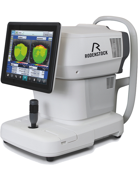
The energy levels in the self setting program can reach 31Jcm2 with high intensity of 4,000W. | Rodenstock Alino Multifunction Refractometer
Tune-up your businessExperience 6 analyses in 1 device: Rodenstock Alino Multifunction Refractometer
Are you ready for something that is all-in-one, timesaving, space-saving and suits your business perfectly?Have a look at how powerful ALINO can be.6 in 1 solution01 RefractionHaving a good starting value for subjective refraction is essential. The QUICK REF MODE supports this even for uncooperative patients.02 KeratometryKAI (Kerato-asymmetry index) & KRI (Kerato-regularity index) give the first hint of possible irregularities on the cornea. This supports evaluation of the best vision correction or best-fitting contact lens.03 TonometryThe very gentle air puff created by a new generation of air-flow technology means that patients more readily accept the need for regular check-ups of the IOP measurement.04 PachymetryCorrecting the IOP by measuring the central corneal thickness is essential in interpreting the intraocular pressure.05 TopographyVarious topographic maps support you in carrying out vision screenings, contact lens fittings and in patient education.06 Dry EyeObserve the customers’ eye health and evaluate the long-term tolerability of contact lenses.Take advantage of the combination of tests:Additional servicesTonometry + PachymetryContact lens fitting andtolerance controlKeratometry + Topography + Dry EyeEye healthcareTopography + Tonometry + Pachymetry + Dry EyeSpecifications Rodenstock Alino Multifunction Refractometer
REFRACTIVE POWER MEASUREMENTSpherical refractive power (S) -30.00 D to + 25.00 D (at VD = 12.0 mm)Cylindrical power (C) 0.00 D to ± 12.50 D (at VD = 12.0 mm)Astigmatic axis (A) 0° to 180°KERATOMETRY MEASUREMENTCorneal curvature radius 5.00 mm to 13.00 mmCorneal astigmatic axis 0° to 180°INTRAOCULAR PRESSURE MEASUREMENTMeasurement range 1 mmHg to 60 mmHG (1 hPa to 80 hPa)PACHYMETRY MEASUREMENTMeasurement range 300 μm to 800 μmTOPOGRAPHY MEASUREMENTCorneal curvature radius 5.50 mm to 10.00 mmCorneal astigmatic axis 0° to 180°AUXILIARY FUNCTIONInterpupillary distance Measurement range: 20 mm to 85 mmCorneal diameter and pupil diameter Measurement range: 1.00 mm to 14.00 mmDry eye observation Blinking Analysis, Tear Meniscus height, Hyperemia, Meibomian glandsDATA MANAGEMENTInternal Database Integrated SD CardPrinter Integrated thermal printerData Output Type 3x USB 2.0, 1x LAN, 1x SD Card-slot, 1x WLANExport Format CSV, XML, JPG, PDFDIMENSIONS & ELECTRIC REQUIREMENTSDimensions WDH 312 × 491 × 450 mmWeight Approx. 23 kgVoltage 100 VAC to 240 VACFrequency 50/60 HzPower consumption 110 VA | Siemens Acuson Sequoia 512 LCD Ultrasound Machine18″ Flat panel LCD Monitor–Progressive Media & Display II Package, Cardiac/OB/Vascular Calc. Package, Signature II Option, AUX, CPS Cardiology, CPS General Cadene Trigg Burst, Calc Data to MO, Card Cadence II Contrast Quant, Cornoary Flow reserve, Dicome Store/Print, DTI (Doppler Tissue Imaging), High HR ECG, MIS/MIF, Modality Worklist Native TEQ, Native TEQ, NTHI (Native Tissue Harmonics), Spectral TEQ, TEQ (Tissue Equalization), VVIFeatures- Shared Service
- Native Patient Specific Imaging
- Four Sight TEE view
- Axius quantitative strain rate imaging
- Cadence contrast pulse sequencing technology
- Native Tissue Harmonic Imaging
- Native TEQ ultrasound technology
- Color doppler, Color angio and b-color
- PW/CW Doppler
- M-Mode
- Folding Flat Panel Display
- DICOM capabilities
Applications- Abdominal
- Breast
- OB/GYN
- Vascular
- Pediatrics
- Cardiology
- Musculoskeletal
- Neonatal
Acuson Sequoia 512 Features:- Imaging: Gray-scale 2-D, M-mode, SST Color Doppler, Solo Spectral Doppler, 3-D Surface Rendering, Native Tissue Harmonic Imaging, Auto Doppler, M-Mode, PW Doppler, Color Doppler, Color Power Doppler
- 19" color CRT monitor
- Native tissue harmonic imaging (NTHI)
- DIMAQ: image management
- Advanced vascular analysis
- Advanced spatial compounding
- Native patient specific imaging
- Mitral valve assessment
- CPS contrast pulse sequencing technology
- Clarifying vascular enhancement technology
- High resolution color flow
- Vascular, and General Imaging calculation packages
- Coherent beamformer technology/spatial compounding
- Space/time resolution control
- Cine and zoom/RES
Compatible Ultrasound Probes and Transducers:- Acuson V5Ms TEE Probe
- Acuson 4C1 Curved Array Probe
- Acuson 6C2 Curved Array Probe
- Acuson 8C4 Curved Array Probe
- Acuson 4V1 Vector Array Probe
- Acuson EV-8C4 Endocavitary Probe
- Acuson EC-10C5 Endocavitary Probe
- Acuson 6L3 Linear Array Probe
- Acuson 8L5T Intraoperative Probe
- Acuson 8L5 Linear Array Probe
- Acuson 9L4 Linear Array Probe
- Acuson 15L8 Linear Array Probe
- Acuson 17L5 Linear Array Probe
The Sequoia 512 with LCD ultrasound machine uniquely minimizes automatic image optimization time by up to 80% to make way for an easier and speedier ultrasound test time as well as allow for a more confident diagnosis each time. As a patient-specific imaging ultrasound, it has a unique matched response capacity for accurate image quality, sharp clinical specificity, and optimized diagnostic outcomes that makes infinitesimal pathologies and other anatomical detail in the most difficult-to-image patients becomes more vivid and visible. | Zeiss Cirrus HD-OCT 4000 OCT Spectral Domain OCT HD
Accessories- The ZEISS Certified Factory Direct Cirrus 4000 HD-OCT also includes the full Accessory Kit
- External Fixation Light
- Test Eye
- Mini Keyboard
- Optical Mouse
- Dust Cover
- Metric 5A/250v Fuse
- 14 Ft. CAT 5e Network Cable
- USB Cable
- IEC 320 to NEMA 12 inch Power Cable
- IEC 320 39in. Power Cable
- Install Disc including Roxio Basic
- Symbol Sheet
- OCT Primer Guide
- Quick Start Guide
HD-OCT Imaging- Methodology: Spectral Domain OCT
- Optical Source: superluminescent diode (SLD), 840 nm
- Optical Power: < 725 μW at the cornea
- Scan Speed: 27,000 A-scans per second
- A-Scan depth: 2.0 mm (in tissue), 1024 points
- Axial resolution: 5 μm (in tissue)
- Transverse resolution: 15 μm (in tissue)
Fundus Imaging- Methodology: Line scanning ophthalmoscope
- Live Fundus Image: During alignment and during OCT scan
- Optical Source: Superluminescent diode (SLD), 750 nm
- Optical Power: < 1.5 mW at the cornea
- Field of View: 36 degrees W x 30 degrees H
- Frame rate: >20 Hz
- Transverse resolution: 25 μm (in tissue)
Iris Imaging- Methodology: CCD Camera
- Resolution: 1280 x 1024
- Live iris image: During alignment
Electrical, Physical and Environmental- Weight: 38kg (83 lbs)
- Dimensions: 65L x 44W x 53H (cm)
- Fixation: Internal, external
- Internal fixation focus adjustment: -20D to +20D (diopters)
- Input devices: Keyboard, mouse
- Electrical rating (115V) Single Phase, 100/120V~ systems:50-60Hz, 5A
- Fuse rating (115V) T 5A 250V
- Electrical rating (230V) Single Phase, 220/240V~ systems:50-60 Hz, 2.5A
- Fuse rating (230V) Fuse rating: T 5A 250V
- Convenience Receptacle output ratings 115V~, 0.5 A Max, 50-60 Hz
- Temperature (transport and storage) -40º to +70º C
- Relative humidity (transport and storage) 10% to 100%, including condensation
- Atmospheric pressure (transport and storage): 500 hPa to 1060 hPa
- Temperature (operation) +10º to +35º C
- Relative humidity (operation) 30% to 75%, excluding condensation
- Atmospheric Pressure (operation) 700 hPa to 1060 hPa
Computer Zeiss Cirrus HD-OCT 4000 OCT Spectral Domain OCT HD- • High performance multi-core processor
- • Internal storage: > 80,000 scans
- • CD-RW, DVD-ROM drive
- • Integrated 15″ color flat panel display.
The Cirrus HD-OCT is a non-contact, high resolution tomographic and biomicroscopic imaging device. It is indicated for in-vivo viewing, axial cross-sectional, and three-dimensional imaging and measurement of anterior and posterior ocular structures, including cornea, retina, retinal nerve fiber layer, ganglion cell plus inner plexiform layer, macula, and optic nerve head. The Cirrus normative databases are quantitative tools for the comparison of retinal nerve fiber layer thickness, macular thickness, ganglion cell plus inner plexiform layer thickness, and optic nerve head measurements to a database of normal subjects. The Cirrus HD-OCT is intended for use as a diagnostic device to aid in the detection and management of ocular diseases including, but not limited to, macular holes, cystoid macular edema, diabetic retinopathy, age-related macular degeneration, and glaucoma. | Carl Zeiss Cirrus HD-OCT 5000 Smart Advanced Topography HDAdvancing Smart OCTfor Anterior Segment, Glaucoma and Retina- Anterior Segment Premier Module
- PanoMap Wide-Field Display
- Smart HD Scans
- En face report
Visualization at the speed of CIRRUSAnalyzing a single pathology from multiple views provides comprehensive insight and analysis of the clinical situation. How this helps you: - Spot small areas of pathology. Tightly spaced B-scans, (either 30 or 47 μm apart), in the cube ensure that small areas of pathology are imaged. For reference, a human hair is about 40-120 μm in diameter.
- Visualize the fovea. Scans that are spaced further apart than in the CIRRUS cube may miss the central fovea.
- Fuel for analysis. Millions of data points from the cube are fed into the Zeiss proprietary algorithms for accurate segmentation, reproducible measurements and registration for change analysis.
- Take the pressure off the operator. As long as the scan is placed in the vicinity of the fovea or optic nerve, the software automatically centers the measurements after the capture.
- See the tissue from different perspectives. View the cube data from all angles, with 3D rendering, OCT fundus images and Advanced Visualization™.
- Future ready. Previously captured CIRRUS cubes can be analyzed using new analyses.
Tracking at the speed of CIRRUSFastTrac™ reduces eye motion artifacts without sacrificing patient throughput with a proprietary scan acquisition strategy, high speed 20 Hz LSO camera, and single-pass alignment scanning
With FastTrac, scan at the highest resolution at the same location at each visit. Assessment at the speed of CIRRUSMeasurement centering with FoveaFinder and AutoCenterOn the macula, the unique FoveaFinder technology ensures the ETDRS and ganglion cell plus inner plexiform layer measurement frameworks are centered on the fovea.AutoCenter™ function automatically centers the 3.4 mm diameter peripapillary RNFL calculation circle around the disc for precise placement and repeatable registration. The placement of the circle is not operator dependent. Accuracy, registration and reproducibility are assured. Manage at the speed of CIRRUS Carl Zeiss Cirrus HD-OCT 5000 Smart Advanced Topography HDGuided Progression Analysis (GPA™) for Assessing RNFL and Optic Disc ChangeGPA™ compares RNFL thickness and optic disc measurements over time and determines if statistically significant change has occurred.Focal Progression Analysis – Up to 6 progression maps are compared to two baselines. Areas of statistically significant change are color-coded yellow when first noted and then red when the change is sustained over consecutive visits.
Trend Analysis with Rate of Change – RNFL thickness values are plotted over time. Trends are calculated and confidence intervals are shaded when rate of change is statistically significant. |




Reviews
There are no reviews yet.