| Description | CIRRUS 4000 delivers high-speed image capture with HD imaging detail and a wider field of view so you can make more informed decisions and spend more time with the patients who need it. | Experience 6 analyses in 1 device: refraction, keratometry, topography, tonometry, pachymetry and dry eye analysis. | CIRRUS 6000 is the next-generation OCT from ZEISS, delivering high-speed image capture with HD imaging detail and a wider field of view so you can make more informed decisions and spend more time with the patients who need it.Learn how you can maximize patient throughput and practice efficiency with ZEISS CIRRUS 6000. | The new tri-functional Autorefractor Keratometer Topcon KR-8000PA Supra combines an autorefractor, an autokeratometer, and a computerized color corneal mapping system all in a single, compact unit! This enables the instrument to provide three times the diagnostic capabilities in one-third the space, at a fraction of the cost of the individual instruments. | RIGHTON RETINOMAX-5 AUTO REFRACTOMETER / KERATOMETER
Automatic AxisCompensation and extended measurement range
Not only does the device let examiner know the cylinder axis angle, but it can
also be automatically adjusted if it is not level. | The Tomey Refractometer Keratometer and Topographer only in 1 unit senior citizens and children can also get the inspection with out any trouble because they dont have to move the seat for three measurements. |
| Content | Carl Zeiss Cirrus HD-OCT 4000
The Cirrus HD-OCT is a non-contact, high resolution tomographic and biomicroscopic imaging device. It is indicated for in-vivo viewing, axial cross-sectional, and three-dimensional imaging and measurement of anterior and posterior ocular structures, including cornea, retina, retinal nerve fiber layer, ganglion cell plus inner plexiform layer, macula, and optic nerve head. The Cirrus normative databases are quantitative tools for the comparison of retinal nerve fiber layer thickness, macular thickness, ganglion cell plus inner plexiform layer thickness, and optic nerve head measurements to a database of normal subjects. The Cirrus HD-OCT is intended for use as a diagnostic device to aid in the detection and management of ocular diseases including, but not limited to, macular holes, cystoid macular edema, diabetic retinopathy, age-related macular degeneration, and glaucoma.The ZEISS Certified Factory Direct Cirrus 4000 HD-OCT also includes the full Accessory Kit
External Fixation Light
Test Eye
Mini Keyboard
Optical Mouse
Dust Cover
Metric 5A/250v Fuse
14 Ft. CAT 5e Network Cable
USB Cable
IEC 320 to NEMA 12 inch Power Cable
IEC 320 39in. Power Cable
Install Disc including Roxio Basic
Symbol Sheet
OCT Primer Guide
Quick Start GuideHD-OCT ImagingMethodology: Spectral Domain OCT
Optical Source: superluminescent diode (SLD), 840 nm
Optical Power: < 725 μW at the cornea
Scan Speed: 27,000 A-scans per second
A-Scan depth: 2.0 mm (in tissue), 1024 points
Axial resolution: 5 μm (in tissue)
Transverse resolution: 15 μm (in tissue)Zeiss Cirrus HD-OCT 4000 OCT Spectral Domain OCT HD Fundus ImagingMethodology: Line scanning ophthalmoscope
Live Fundus Image: During alignment and during OCT scan
Optical Source: Superluminescent diode (SLD), 750 nm
Optical Power: < 1.5 mW at the cornea
Field of View: 36 degrees W x 30 degrees H
Frame rate: >20 Hz
Transverse resolution: 25 μm (in tissue)Carl Zeiss Cirrus HD-OCT 4000 Iris ImagingMethodology: CCD Camera
Resolution: 1280 x 1024
Live iris image: During alignmentElectrical, Physical and EnvironmentalWeight: 38kg (83 lbs)
Dimensions: 65L x 44W x 53H (cm)
Fixation: Internal, external
Internal fixation focus adjustment: -20D to +20D (diopters)
Input devices: Keyboard, mouse
Electrical rating (115V) Single Phase, 100/120V~ systems:50-60Hz, 5A
Fuse rating (115V) T 5A 250V
Electrical rating (230V) Single Phase, 220/240V~ systems:50-60 Hz, 2.5A
Fuse rating (230V) Fuse rating: T 5A 250V
Convenience Receptacle output ratings 115V~, 0.5 A Max, 50-60 Hz
Temperature (transport and storage) -40º to +70º C
Relative humidity (transport and storage) 10% to 100%, including condensation
Atmospheric pressure (transport and storage): 500 hPa to 1060 hPa
Temperature (operation) +10º to +35º C
Relative humidity (operation) 30% to 75%, excluding condensation
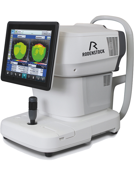
Atmospheric Pressure (operation) 700 hPa to 1060 hPa | Rodenstock Alino Multifunction Refractometer
Tune-up your businessExperience 6 analyses in 1 device: Rodenstock Alino Multifunction Refractometer
Are you ready for something that is all-in-one, timesaving, space-saving and suits your business perfectly?Have a look at how powerful ALINO can be.6 in 1 solution01 RefractionHaving a good starting value for subjective refraction is essential. The QUICK REF MODE supports this even for uncooperative patients.02 KeratometryKAI (Kerato-asymmetry index) & KRI (Kerato-regularity index) give the first hint of possible irregularities on the cornea. This supports evaluation of the best vision correction or best-fitting contact lens.03 TonometryThe very gentle air puff created by a new generation of air-flow technology means that patients more readily accept the need for regular check-ups of the IOP measurement.04 PachymetryCorrecting the IOP by measuring the central corneal thickness is essential in interpreting the intraocular pressure.05 TopographyVarious topographic maps support you in carrying out vision screenings, contact lens fittings and in patient education.06 Dry EyeObserve the customers’ eye health and evaluate the long-term tolerability of contact lenses.Take advantage of the combination of tests:Additional servicesTonometry + PachymetryContact lens fitting andtolerance controlKeratometry + Topography + Dry EyeEye healthcareTopography + Tonometry + Pachymetry + Dry EyeSpecifications Rodenstock Alino Multifunction Refractometer
REFRACTIVE POWER MEASUREMENTSpherical refractive power (S) -30.00 D to + 25.00 D (at VD = 12.0 mm)Cylindrical power (C) 0.00 D to ± 12.50 D (at VD = 12.0 mm)Astigmatic axis (A) 0° to 180°KERATOMETRY MEASUREMENTCorneal curvature radius 5.00 mm to 13.00 mmCorneal astigmatic axis 0° to 180°INTRAOCULAR PRESSURE MEASUREMENTMeasurement range 1 mmHg to 60 mmHG (1 hPa to 80 hPa)PACHYMETRY MEASUREMENTMeasurement range 300 μm to 800 μmTOPOGRAPHY MEASUREMENTCorneal curvature radius 5.50 mm to 10.00 mmCorneal astigmatic axis 0° to 180°AUXILIARY FUNCTIONInterpupillary distance Measurement range: 20 mm to 85 mmCorneal diameter and pupil diameter Measurement range: 1.00 mm to 14.00 mmDry eye observation Blinking Analysis, Tear Meniscus height, Hyperemia, Meibomian glandsDATA MANAGEMENTInternal Database Integrated SD CardPrinter Integrated thermal printerData Output Type 3x USB 2.0, 1x LAN, 1x SD Card-slot, 1x WLANExport Format CSV, XML, JPG, PDFDIMENSIONS & ELECTRIC REQUIREMENTSDimensions WDH 312 × 491 × 450 mmWeight Approx. 23 kgVoltage 100 VAC to 240 VACFrequency 50/60 HzPower consumption 110 VA | New ZEISS CIRRUS 6000 – 100 kHz OCT Imaging SystemAt 100,000 scans per second, ZEISS CIRRUS 6000 enables clinicians to image a larger field of view up to 12mm in a single scan. It also captures high-defintion (HD) OCT and OCT Angiography (OCTA) scans, revealing the finer microvascular details of the retina and providing more insight into your patient’s condition. AngioPlexOCT Angiography from ZEISS ushers in a new era of eye care with non-invasive imaging of retinal microvasculature—taking glaucoma and retinal disease management and treatment planning to the next level. By offering the industry’s most comprehensive tools for assessing and analyzing a range of pathologies, ZEISS provides a complete OCT Angiography (OCTA) solution. CIRRUS 6000 is the latest addition to an already robust portfolio of OCT devices that are designed to meet the needs of all types and levels of practices and/or clinicians. It offers customers high-speed and high-throughput to help manage their large patient volume faster, all while improving imaging and image quality. “This system was designed with the patient in mind, enabling doctors to continue accessing their patient data from CIRRUS devices that are crucial to effective long-term patient care,” said Jim Mazzo, Global President of Ophthalmic Devices at Carl Zeiss Meditec. “We are committed to our customers and believe this will become an essential asset to their practice.”The 100kHz speed of the CIRRUS 6000 allows clinicians to scan patients faster, with increased efficiency and improved imaging detail. Additional features include: - Wider and deeper OCT/OCTA scans with 12x12mm OCTA and B-scan depth up to 2.9mm
- HD AngioPlex scan is a new scan type that provides more detail
- New workflow protocols to aid in increasing efficiency
- Patient-friendly “Wellness report” to help educate the patient
According to retina and vitreoretinal physician and surgeon Gregg T. Kokame, MD, of Retina Consultants of Hawaii, the new ZEISS CIRRUS 6000 is extremely fast and impressive. “With the new scan acquisition time of 100,000 per second I can scan and assess my patients much quicker, giving me the details I need with every image,” says Kokame.“ZEISS is dedicated to advancing eye care through development of new and innovative solutions, like the CIRRUS 6000,” said Dr. Ludwin Monz, President and CEO of Carl Zeiss Meditec AG. “With our comprehensive diagnostics portfolio, we will continue to create better vision standards to benefit patients today and in the future.” | Topcon KR8000PA Autorefractor KeratometerBy implementing Topcon’s Autorefractor Keratometer Topcon KR-8000PA Supraexclusive rotary prism technology for the refraction portion of the measurement, the Autorefractor Keratometer Topcon KR-8000PA Supra provides objective data that is unmatched in accuracy and reliability. Auto tracking and auto measurement enhance the ease of operation. The Autorefractor Keratometer Topcon KR-8000PA Supra unit also incorporates ten placido rings for enhanced testing precision, providing corneal mapping measurements out to 10mm! |
Features for Autorefractor Keratometer Topcon KR-8000PA SupraTopcon Color Mapping 32 SoftwareThis versatile 32-bit color mapping software system enhances clinical evaluations with the output of highly accurate, full color corneal maps. The system offers an enlarged color map, tangential axial maps, sagittal maps, refractive and keratometric data, and peripheral keratometric maps. It provides early phase detection of subclinical keratoconus as well as pre and post-op evaluation. The software also features a new image overlay function, as well as a Multi-Map and Difference Map View, which display multiple image maps for easy comparison. The color mapping system is an excellent patient education tool and is designed for use with the Topcon KR-7000P or KR-8000PA Auto Refractor/Keratometer. Windows® 95/98/NT Compatible.
The color mapping software also features a contact lens fitting module with adjustable parameters to view the contact lens fit on screen.The Autorefractor Keratometer Topcon KR-8000PA Supra System Benefits Include:Topcon KR8000PA Autorefractor Keratometer- Allows Three Functions at One Station:
– Refraction
– Keratometry
– Corneal Mapping
- Contact Lens Fitting Utility
- Early Phase Detection of Subclinical Keratoconus
- Direct Link to CV System
The new tri-functional Autorefractor Keratometer Topcon KR-8000PA Supra combines an autorefractor, an autokeratometer, and a computerized color corneal mapping system all in a single, compact unit! This enables the instrument to provide three times the diagnostic capabilities in one-third the space, at a fraction of the cost of the individual instruments. | Righton Retinomax 5 Auto Refractor
The Retinomax Series, the world’s leading handheld autorefractor and autorefractor/keratometer, delivers wide measurement range on par with table type refr, now boasts greater mobility, stability and accuracy. The Retinomax series is widely used as a global standard device for pediatric, screening, medical care in remote areas, disaster area and wide range of ophthalmic scene.FEATURES- View Finder – With view finder observation, no limit of operator age. Diopter adjustable range is +/-8D the same as Retinomax3. View finder arm angle is changeable of 0-135 degrees, making measurement easy regardless of patient’s position or posture. Adjustable diopter +/- 8D.
- Wider Eye Piece – Eye piece relief is 48mm from 32.2mm so that enable to look inside image while even looking at outside. No need to stick eye to the eyepiece all the time.
- Improved battery life – At 180 minutes, the battery capacity is now twice that of conventional models
- Automatic Axis Compensation and Extended Measurement Range – Not only does the device let examiner know the cylinder axis angle, but it can also be automatically adjusted if it is not level
- New Child Mode – While the measurement is being taken, a melody plays continually to keep children’s attention. A constantly changing color display, both on the outside and inside target the device, also keeps children involved during the process.
- Light weight and easy-to-hold grip
View Finder Righton Retinomax 5 Auto RefractorWith view finder observation, no limit of operator age. Diopter adjustable rangeis +/-8D the same as Retinomax3. View finder arm angle is changeable of 0-135degrees, making measurement easy regardless of patient’s position or posture.Adjustable diopter +/- 8D.Improved battery lifeAt 180 minutes, the battery capacity is now twice that of conventional modelsWider Eye PieceEye piece relief is 48mm from 32.2mm so that enable to
look inside image while even looking at outside. No need
to stick eye to the eyepiece all the time.Focus AssistLets you know the exact focusing point with
the following color-coding system.New Child ModeWhile the measurement is being taken, a
melody plays continually to keep children’s
attention. A constantly changing color
display, both on the outside and inside of
the device, also keeps children involved
during the process.Automatic AxisCompensation and extended measurement range
Not only does the device let examiner know the cylinder axis angle, but it can
also be automatically adjusted if it is not level. | Tomey RT-7000 Autorefractor Keratometer Topographer
-Three Functions in One Instrument-Easy & Speedy Touch Screen Alignment-6.4 inches color TFT LCD-Dual CCD technology for Refractometry-IOL/ Cataract Mode-New incites for Keratometry KAI, KRI-Auto Alignment / Auto Shot of TopographyCorneal Topography– Auto Alignment / Auto ShotAny one can easily capture measurement with Auto Alignment and Auto Shot. The Measurement variation is significantly reduced no matter what the skill level is of the operator.CL Fitting Simulation-A pseudo-fluorescent pattern of the contact lens selected from the built-in data base can be confirmed and the fitting can be stimulated before the patient wears the trial lens.Corneal Eccentricity Index-The index of CEI (Corneal Eccentricity Index) indicates the ratio of flattering in percentage from the central cornea to the periphery.Three Function in One Instrument– Refractometer Keratometer and Topographer only in 1 unit senior citizens and children can also get the inspection with out any trouble because they don’t have to move the seat for three measurements. Switch from Ref-Kerato Mode to Corneal Topography Mode with only ONE TOUCH. The light come appears with only one button from the measuring head and the Ref-Keratometer is transformed to the topographer.Refractometry– Dual CCD technology for RefractometryTwo CCD cameras are used to capture images for observation and for measurement while providing highly accurate measurement data.Keratometory– New Indices for KeratometryThe Corneal Irregular astigmatism display function is installed to expand the possibility of Keratometer. This is the new function to measure the level of corneal irregular astigmatism which was difficult in the past.SPECIFICATIONSMeasurmentRangesRefraction MeasuremntSphere -25.00D to ±22.00DIVD=12.0mmCylinder 0D to ± 10.00DIVD=12.0mmAxis 0 to 180Corneal Curvature Measurement 5.00mm to 11.00mmMeasurement Zone 3mm R:8.00mmCorneal MappingDisplay Range 9 to 100 D-Normal Mode1.0 to 8.0mm (R:8.00mm)-Special Mode0.9 to 7.0 mm (R:8.00mm)Measurement TimeREF 0.2 SecondsKRT 0.1 SecondsMinimum Pupil Diameter 2.2mmMinimum Measurment Step Refraction MeasurementSphere 0.01DCylinder 0.01DAxis 1Corneal Curvature Measurement 0.01mmPD (Pupil Distance) 50mm to 86mmRecording Built-In Thermal PrinterVertex Distance 0mm, 12.0mm, 13.5mm, 14.0mm, 15.5mm, 16.0mmExterior Output (AN port / 4USB ports)Dimensions Tomey RT-7000 Autorefractor Keratometer TopographerHigh 466mmm or 18 InchesWidth 307mm or 12 InchesDepth 490mm or 19 InchesWeight Approx 20,000Kg / 44.4 PoundsDisplay 6.4 Inches color liquid crystalPower SupplyVoltage AC 100 to 240VFrequency 50/60 HzConsumption Power 120VA to 150VATemperature +10C to + 40CDisplay 30% to 75% |




Reviews
There are no reviews yet.