View cart “Sirona Apollo DI Intra Oral Scanner For Sale” has been added to your cart.
-46%
Availability: In Stock
SOTA Clio Digital X-Ray Sensor Size 2 For Sale
$5,200.00$2,800.00
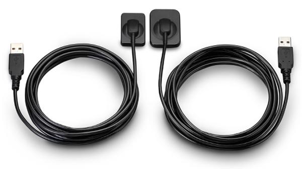
The Clio Digital X-ray Sensor is equipped with a patented, Kevlar reinforced cable to provide maximum durability. Unlike other products, the USB cable of the Clio is fully replaceable if damaged. Constructed with special shock absorbers inside the sensor housing to protect components from exterior damage, the Clio is also one of the few fully water submersible sensors, sealed with a unique rubber polymer interior housing.
Other people want this. 15 people have this in their carts right now.
The Clio Digital Dental X-Ray Sensors have Next Generation CMOS technology, fiber optic sensors create 20+ actual line pairs per mm (26.3 theoretical line pairs per mm).With our Cliosoft Imaging Software, we can gaurantee our Clio Digital Dental X-Ray images match up or surpass any competing system. We make this promise based on feedback from actual dentists.Rounded edges allow for maximum patient comfort. Ideal intraoral sensor width and thinness in two sizes for pedo and adult.
The Clio Digital X-Ray Sensors maximize image area ratio, minimizing cutoff space to mimic the size of traditional x-ray film. Next Generation CMOS technology fiber optic sensors create 20+ actual line pairs per mm (26.3 theoretical). With our user-friendly, tablet and PC-ready cliosoft Digitial Imaging Software, we guarantee the highest quality images in the industry.
Features:
– All new Direct Integration Technology
– Thin sensor hardware with rounded corners for maximum comfort
– Direct USB cable with no docking station
– Repairable USB cable for longevity of sensors
– Fiber optic plate sensors for low-noise ratio
– Largest active area for maximum coverage
– Fully waterproof sensor head with sealed cable and housing design
– USB connector: USB 2.0, USB 3.0
– Sensor Technology
– Next Generation CMOS Fiber optic Sensors
– Maximum Gray Levels – 4096 (12-bit)
Specifications:
– External Dimensions: 30.49 x 42.80 mm
– Active Area: 25.99 x 36.02 mm
– Sensor Plate Thickness: 5.41 mm
– Sensor Cable Length: 3 meters
– Image Resolution: 26.3 lp/mm theoretical 20 lp/mm actual
– Image Size: 1896 x 1368 pixels
– Image Pixel Size: 19×19 microns
The Clio Digital X-ray Sensor is equipped with a patented, Kevlar reinforced cable to provide maximum durability. Unlike other products, the USB cable of the Clio is fully replaceable if damaged. Constructed with special shock absorbers inside the sensor housing to protect components from exterior damage, the Clio is also one of the few fully water submersible sensors, sealed with a unique rubber polymer interior housing.
Reviews (0)
Reviews
There are no reviews yet.
Be the first to review “SOTA Clio Digital X-Ray Sensor Size 2 For Sale” Cancel reply
The Clio Digital X-ray Sensor is equipped with a patented, Kevlar reinforced cable to provide maximum durability. Unlike other products, the USB cable of the Clio is fully replaceable if damaged. Constructed with special shock absorbers inside the sensor housing to protect components from exterior damage, the Clio is also one of the few fully water submersible sensors, sealed with a unique rubber polymer interior housing.
Schick Technologies has brought digital panoramic imaging to a whole new level of affordability and simplicity.
Benefits:
Faster Scan Speed
Superb Image Quality
Light Guidance System
Portable for Easy Sharing
Lightweight Hand Piece
iTero HD2.9 simply allows to digitally scan a prepared tooth and wirelessly send this information directly to the dental laboratory for fabrication of a CAD/CAM crown without the gooey mess.
Are you paying too much for digital X-ray sensors and warranties? Consider QuickRay from Video Dental Concepts! A dental imaging leader and innovator since 1989, VDC has provided over 7,000 dental professionals with the best diagnostic imaging values and personalized support for over 20 years.
The Waterlase MD features Hydro- Photonics?a combination of YSGG laser energy and water?which allows the clinician to perform a wide range of conventional procedures with less anesthetic. Known for its precision and accuracy, the MD enables treatment of targeted areas of both tooth structure and soft tissue while maintaining the structural integrity of the tooth and leaving surrounding areas unaffected.
Content
SOTA Clio Digital X-Ray Sensor Size 2
SOTA Clio Digital X-Ray Sensor Size 2The Clio Digital Dental X-Ray Sensors have Next Generation CMOS technology, fiber optic sensors create 20+ actual line pairs per mm (26.3 theoretical line pairs per mm).With our Cliosoft Imaging Software, we can gaurantee our Clio Digital Dental X-Ray images match up or surpass any competing system. We make this promise based on feedback from actual dentists.Rounded edges allow for maximum patient comfort. Ideal intraoral sensor width and thinness in two sizes for pedo and adult.The Clio Digital X-Ray Sensors maximize image area ratio, minimizing cutoff space to mimic the size of traditional x-ray film. Next Generation CMOS technology fiber optic sensors create 20+ actual line pairs per mm (26.3 theoretical). With our user-friendly, tablet and PC-ready cliosoft Digitial Imaging Software, we guarantee the highest quality images in the industry.Features:– All new Direct Integration Technology
– Thin sensor hardware with rounded corners for maximum comfort
– Direct USB cable with no docking station
– Repairable USB cable for longevity of sensors
– Fiber optic plate sensors for low-noise ratio
– Largest active area for maximum coverage
– Fully waterproof sensor head with sealed cable and housing design
– USB connector: USB 2.0, USB 3.0
– Sensor Technology
– Next Generation CMOS Fiber optic Sensors
– Maximum Gray Levels – 4096 (12-bit)Specifications:– External Dimensions: 30.49 x 42.80 mm
– Active Area: 25.99 x 36.02 mm
– Sensor Plate Thickness: 5.41 mm
– Sensor Cable Length: 3 meters
– Image Resolution: 26.3 lp/mm theoretical 20 lp/mm actual
– Image Size: 1896 x 1368 pixels
– Image Pixel Size: 19×19 microns
The Clio Digital X-ray Sensor is equipped with a patented, Kevlar reinforced cable to provide maximum durability. Unlike other products, the USB cable of the Clio is fully replaceable if damaged. Constructed with special shock absorbers inside the sensor housing to protect components from exterior damage, the Clio is also one of the few fully water submersible sensors, sealed with a unique rubber polymer interior housing.
Schick CDR PanX Laser
FeaturesCDRPanX features proven Schick CDR digital imaging technology and renders crisp, clinically accurate images
Only slightly more expensive than most film-based systems, CDRPanX eliminates costly panoramic film and processing – increasing your economic efficiency
Eight different imaging modes and three laser alignment beams for optimal exposures
Superior vertical travel allows it to be used with patients of any height, including those in wheelchairs
Patient Positioning: 3 laser alignment beams (mid-sagittal, Frankfort, focal trough) bite guide, chin rest, optional temple support
SpecificationsGenerator: Multipulse DC
X-Ray Tube: OCX105
Focal spot size: 0.5 IEC 336
Minimum total filtration: 2.5 mm Al
Anode Voltage: 60-86kV (2kV increments)
Anode Current: 4, 5, 6.3, 8, and 10mA
Exposure time: 19s – adult panoramic
Power Supply: 115V, 50/60Hz
Fusing: T8A
Weight: 474lbs
Electrical Safety Classification: Class 1, type B
Imaging modes (Exposure times): adult (19s), child (15s), left half (10s), right half (10s), anterior (8s), TMJ opened and closed (4x4s), frontal sinuses (8s)
Patient Positioning: 3 laser alignment beams (mid-sagittal, Frankfort, focal trough) bite guide, chin rest, optional temple supportFeatures
CDRPanX features proven Schick CDR digital imaging technology and renders crisp, clinically accurate images
Only slightly more expensive than most film-based systems, CDRPanX eliminates costly panoramic film and processing – increasing your economic efficiency
Eight different imaging modes and three laser alignment beams for optimal exposures
Superior vertical travel allows it to be used with patients of any height, including those in wheelchairs
Carestream CS 3600 Intraoral ScannerAcquire true color:With the CS 3600 Intraoral Scanner, practitioners can access 2D and 3D images that can be used with CS Restore software to design restorations within their practice. Colored 3D scans allow practitioners to easily define the margin lines and identify the differences between natural tooth structure and existing restorations. This makes restoration planning more accurate for practitioners.No external heater, powder or trolley system needed:The CS 3600 does not require you to spray powder of liquid over the patient’s teeth or gingival tissue before scanning. It features high-angulation scanning of up to 45 degrees and to a depth from -2 to +13m.Features a unique light guidance systemYet another feature of the CS 3600 that assists it in the capture of the data during the image acquisition process. This feature allows the user to place more focus on the patient’s mouth rather than the monitor.This device is easily shared:The CS 3600 can be easily shared between operatories because there is no heavy trolley to push around. Additionally, the intraoral scanner connects via USB 2 cable* to any PC workstation or laptop. * Windows 7, 32-bit minimum; 64-bit recommendedEasy to use and hold:The CS 3600’s sleek, lightweight design (295 grams) makes it comfortable and easy for users to navigate the intraoral scanner around patients’ mouths, especially during full arch acquisitions.Benefits:Carestream CS 3600 Intraoral Scanner
Faster Scan Speed
Superb Image Quality
Light Guidance System
Portable for Easy Sharing
Lightweight Hand Piece
Features
High Speed Continuous Scanning
Fast, Simple and Smooth user experience
Freefill Missing Scan Data at your Leisure
Scan History allows you to remove excess scanned tissue for more refined final digital impression
Three Workflows
user friendly interface
Autoclavable reusable tips in two interchangeable styles
iTero HD2.9 simply allows to digitally scan a prepared tooth and wirelessly send this information directly to the dental laboratory for fabrication of a CAD/CAM crown – without the gooey mess. The wireless transmission saves shipping time and eliminates distortion that can often be found with traditional stone modelsThe digital scan is, quite literally, digitally perfect. We then deliver the digital scan to our lab where a precision fitting restoration is created.iTero HD2.9 system eliminated the uncomfortable patient experience of traditional impressions, utilizes advanced state of the art digital technology, and provides with superior fitting restorations with little or no adjustmentsiTero Intra-Oral Scanner
Model: HD2.9
2014 Date of Mfg.
Brochure/Video: Click Here
Limited Use
Includes:
iTero Software (pre-loaded)
90 Day Parts Warranty
User Manual(s)
Free Standard Shipping (Commercial USA Addresses)
Its ergonomic design comes with safe docking station. It has thin fiber optic cable which enables quick data transfer from the wand to the cart. This optimized cable length assists various cart positions in the operatory.Orthodontic treatment is more than just straightening teeth; it’s about ensuring your teeth and jaws are in alignment. To achieve this, we need to get an accurate look at where your teeth are positioned. To do this, we use the iTero Intraoral digital scanner.A good impressionIn the orthodontic and dental industries, ‘impressions’ is the term we use for getting a good picture of where your teeth currently sit. These impressions were previously taken using a type of dental cement. Pasted on your teeth, patients were required to lie for a few minutes while the cement-like material set. The dental professional could then remove the set material which had formed an ‘impression’ of your teeth.This process was messy, rather unpleasant and not very comfortable. Fortunately, that method is a thing of the past. These days, we use scanning technology and at L&L Orthodontics, our technology of choice is the iTero Intraoral digital scanner.
What is the iTero scanner?
The iTero scanner is new 3D orthodontic scanning technology. Using the latest developments in intraoral scanning, the iTero scanner accurately scans your teeth as part of your orthodontic assessment. The iTero scanner is made up of a small, wand-like device which is used to scan your teeth and jaws. The data it is collecting then appears on the computer screen, allowing you to see the ‘impressions’ the scanner is taking.The iTero scanner works in with Invisalign®* treatment and lets you see what your teeth will look like post-treatment, even before you’ve started. Before we recommend a course of treatment for you, we’ll take impressions of your teeth using the iTero scanner to ensure we are making our recommendation on accurate information.To find out more about the iTero scanner and other orthodontic technology we use here at L&L Orthodontics, book your specialist consultation today.
QuickRay USB Size 1 And 2 X-Ray Sensor
QuickRay USB is a breakthrough in digital X-rays. The embedded electronics are located in the sensor case so there is no interface box! Simply connect the sensor cable via its USB connector to your computer and go! QuickRay USB is currently compatible with these Apteryx based software systems: XVa3, XVLite, Cliniview, Clinitouch, XDR Imaging, MPDX-DMD, Lightyear, DentiMax, ProfSuni, Dr. Suni, Sigma, AFP Digital and Belmont.Great value QuickRay USB digital X-ray sensors connect directly to USB— no interface box needed!Are you paying too much for digital X-ray sensors and warranties? Consider QuickRay from Video Dental Concepts! A dental imaging leader and innovator since 1989, VDC has provided over 7,000 dental professionals with the best diagnostic imaging values and personalized support for over 20 years.QuickRay USB Size 1 And 2 X-Ray Sensor For SaleQuickRay USB is a breakthrough in digital X-rays. The embedded electronics are located in the sensor case so there is no interface box! Simply connect the sensor cable via its USB connector to your computer and go! QuickRay USB is currently compatible with these Apteryx based software systems: XVa3, XVLite, Cliniview, Clinitouch, XDR Imaging, MPDX-DMD, Lightyear, DentiMax, ProfSuni, Dr. Suni, Sigma, AFP Digital and Belmont.The QuickRay sensor gives you the highest resolution and clarity! The thin (less than 1/4″) intraoral sensors include a CMOS image sensor chip, CsI Scintillator, electronic circuits, shielding foils and shock absorbers. Tough polyamide housings have durable replaceable polyurethane cables with ergonomically placed connectors. Their rounded corners and smooth edges optimize patient comfort.
And QuickRay is the fastest sensor on the market! View X-rays in USB 2.0 High Speed Mode in only 3 seconds. Operate in USB 2.0 Full Speed Mode, if necessary.Every sensor is covered by the VDC two year warranty with no annual fees. Technical support is available for an additional fee on an annual basis. Optional pro rata warranty is available from VDC after purchase. All VDC products are FDA registered.
Biolase Waterlase MDX All Tissue LaserThe MDX delivers 300 mJ of energy at 8 W, and it can easily be upgraded to a 450 mJ (9 W) model for 50% faster cutting as clinical skills improve and applications expand (see ?MDX Specs?). This next-generation of the best-selling WaterLase MD features improvements to the laser engine and delivery system ergonomics, and it enables rapid hardtissue cutting speed with unprecedented affordability.Time-Tested Technology Biolase Waterlase MDX All Tissue LaserThe MDX is built on the technologies and reputations of its predecessors, the BIOLASE WaterLase MD and WaterLase iPlus all-tissue lasers.The Waterlase MD features Hydro- Photonics?a combination of YSGG laser energy and water?which allows the clinician to perform a wide range of conventional procedures with less anesthetic. Known for its precision and accuracy, the MD enables treatment of targeted areas of both tooth structure and soft tissue while maintaining the structural integrity of the tooth and leaving surrounding areas unaffected.The Waterlase MD became the company?s flagship product, breaking new ground in the areas of cutting speed, user interface, and ergonomic design. In 2011 that ?flagship? designation was overtaken by the company?s new WaterLase iPlus, an even faster, more powerful machine. It delivers up to 10 W of power at 600 mJ, which makes it the most powerful laser on the global market at the short pulse length of 50 ?s, all through a highly intuitive preprogrammed user interface for everything from routine restorative cases to specialty periodontal, cosmetic, and endodontic procedures.The iPlus features Biolase?s 2780 nm YSGG technology with the versatility of a dual-wavelength configuration; namely, an integrated iLase 940 nm diode laser to handle unexpected soft-tissue cases, better bleeding control, and the potential for temporary pain relief and teeth whitening.
As anticipated, the iPlus proved beneficial for experienced laser dentists who had been anxiously awaiting a machine that would deliver more power, control, and versatility. However, the new laser also provided many of the ?uninitiated? with an easier-than-expected, highly intuitive introduction to all-tissue laser dentistry. The MDX takes this one step further, with a price point that puts the technology within reach of nearly any dentist.
No More Excuses: Affordable Pricing, Substantial ROIThe perception of all-tissue laser technology as inaccessible to most dentists is beginning to dissipate. Even so, some dentists remain unconvinced.The reality, however, is that BIOLASE?s positioning of the MDX as an entry-level product, starting under $40,000, has made the technology much more affordable. Combining better and more efficient manufacturing with strategic sourcing of components results in pricing that most clinicians will view as extremely reasonable. In addition, the initial investment will be recouped with a substantial ROI, which can be realized via 3 main areas of application.1. For hard-tissue restorative dentistry? the ?bread and butter? of general practices?the MDX will enable faster, more efficient and, in most cases, virtually painless restorations for all cavity classes in both adults and children.2. The MDX allows dentists to retain patients who may have traditionally been referred out for osseous crown lengthening. Using the MDX, patients reap the benefits of considerably more comfortable soft-tissue procedures without the inconvenience of a referral, while the practice reaps the benefits of a happier and more loyal patient base.3. MDX provides an effective and efficient gateway into periodontics. Specifically, the MDX promotes deep pocket therapy with new attachment and is especially effective for minimally invasive removal of both subgingival inflamed tissue and calculus in patients with moderate to severe periodontal disease.Another DimensionBIOLASE?s comprehensive in-office and online training and support promise to render the learning curve extremely manageable. And, as intimidation is replaced by both comfort and confidence, clinicians will find that all-tissue laser technology will add a new and profitable dimension to their practices.




Reviews
There are no reviews yet.