| Description | Designed especially for general dentists, the Sirona Orthophos XG3 combines top-tier image quality and exceptional ease-of-use in an affordable, digital panoramic x-ray solution. | Sirona offers your lab greater freedom in terms of indications, materials and for processing third-party CAD/CAM data. The open 5-axis milling and grinding machine inLab MC X5 is designed specifically to meet dental lab requirements for cost-efficient production today and in the future. | Sirona Galileos Dental Cone Beam Imaging System The system captures images using the lowest possible radiation dose and is designed to provide high-quality images for implant planning, treatment guide design, jaw function analysis and more. | The Clio Digital X-ray Sensor is equipped with a patented, Kevlar reinforced cable to provide maximum durability. Unlike other products, the USB cable of the Clio is fully replaceable if damaged. Constructed with special shock absorbers inside the sensor housing to protect components from exterior damage, the Clio is also one of the few fully water submersible sensors, sealed with a unique rubber polymer interior housing. | The inEos X5 can be used to scan impressions and models of all or part of the jaw manually or automatically. The new operating concept increases flexibility for the dental technician so that the scanning process can be optimally integrated into the workflow of the laboratory. While the manual scanning mode saves time for simpler operations, the fully automatic scanning mode has its benefits especially with regard to extensive operations | The affordable Gendex 9200 DDE dental radiography system offers standard panoramic and cephalometric x-ray projections, as well as a wide range of advanced programs to support most dental imaging and exams. |
| Content | SIRONA XG3 Digital Panoramic X-Ray
The ORTHOPHOS XG 3 is designed for the general practice dentist who would like to work with a basic X-ray system. The XG 3 offers you an outstanding price-performance ratio and provides the following benefits:- High image quality
- Extremely simple operation
- Solid workmanship
Accurate positioningThe ORTHOPHOS XG 3 uses proven, logical Sirona principles for exceptional imaging:- Exact, immediate positioning of the anterior teeth in the focal layer
- Effective, comfortable patient stabilisation with forehead and temple supports to prevent motion blurring
Proven TechnologyThe advanced technology from the XG product family enables the ORTHOPHOS XG 3 to produce high quality diagnostic images.
The benefits include:- Specific focal layers and orbital paths for anatomically optimised exposures
- Very detailed display of the anterior tooth region through automatic kV adaption as X-ray beam passes through spinal region
- 16 bit image acquisition technology provides more gray scales
- SIDEXIS XG imaging software with useful analysis tools
Features:SIRONA XG3 Digital Panoramic X-Ray- High-quality, digital panoramic and TMJ imaging power with advanced patient communication tools
- Outstanding price-performance ratio for seamless transition from film to direct digital imaging
- Easy operation with the Multipad; effective, three-point patient stabilization for sharp images
- Direct network capability for exceptional connectivity
SIRONA ORTHOPHOS XG 3The horizontally and vertically reduced pediatric panoramic program offers outstanding image quality with the lowest dose. Ideal for practices that treat a lot of children. The unit’s easy operation and seamless integration into your practice’s daily routines will save you valuable time. The high image quality and proven Sidexis software guarantee that you will come to a safe diagnosis quickly and easily.Quality from the very startHigh quality. Durable. Safe: the Orthophos XG 3 X-ray unit is the perfect basic equipment for general dentists. The unit’s easy operation and seamless integration into your practice’s daily routines will save you valuable time. The high image quality and proven Sidexis software guarantees that you will reach a safe diagnosis quickly and easily.The positioning can be very easily reproduced since image parameters such as the bite block height are saved together with the image data. A bitewing program is available, which helps in special cases and patient situations where intraoral images cannot be used and a shorter panoramic program with a reduced dose, which is suitable for treating children, is also available. |
The new freedom in detailEvery aspect of inLab MC X5 has been designed to address the CAD/CAM needs encountered in dental labs. Intensive technical and technological exchange with many dental technicians has led to the creation of a production machine fulfilling practically all requirements. For the reliable, independent production of dental prostheses.OpeninLab MC X5 is an open production unit. It is the perfect complement to the inLab components inEos X5 and inLab software, but it can also be used to process other STL restoration data. 1 With its own CAM module, the machine can be connected to a wide range of other CAD systems at no additional cost. DesignThe new inLab MC X5 has more than just outstanding functionality and simple operation: its extremely stable construction and modern design are also highly attractive. Its compact, elegant shape will be a real eye catcher in your lab and complements perfectly the award-winning inEos X5 extraoral scanner.Great variety of materialsinLab MC X5 is designed as a universal production unit for processing zirconium oxide, polymers, composites, wax, titanium and metals 2 as well as glass and hybrid ceramics. inLab MC X5 can be used for wet, dry, or dry/wet combination production during the same machining operation depending on material and indication.Sirona has drawn upon its 30 years of experience in the wet processing of glass ceramics to create the ultimate machine for the wet grinding of fully anatomical restorations from high-strength monolithic materials.Switching between wet and dry production e.g. from glass ceramics to zirconium oxide is fast and uncomplicated.inLab MC X5 – efficient and flexible inLab MC X5 is a universal 5-axis production unit for wet and dry production of blocks and disks. It was designed to meet dental requirements in labs and focus on economic and effective production processes.You benefit from a wide range of materials and can select from the high-quality materials of Dentsply Sirona and its CAD/CAM material partners for which the production processes of inLab MC X5 are optimized. | SIRONA GALILEOS COMFORT PLUS
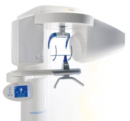
GALILEOS Comfort Plus is an advanced CBCT that provides seamless work flow integration. With an optional HD mode and a 14 second scan, the GALIELOS offers clinicians and specialists numerous options for diagnosis, treatment and patient consultation with superior image quailtiy and low radiation dose. It includes Integrated Implantology and FaceScan, which helps patients better understand and accept treatment recommendations. SICAT Function enables diagnosis and treatment of TMD, and SICAT Air (coming soon) software allows you to anyalze airways.Features:High-Quality Images:
This dental imaging system is able to position itsef and capture a superior quality of images with each scan, ultimately ensuring that you are receiving the detailed images you need, every time.Panoramic:
This imaging system provides crisp panoramic images to your practice with sensors built for high-quality 2D images. With multiple modes, it’s meant to give you the images you need.3D Conebeam:
Equipped with a high-quality, 3D sensor, this machine is capable of producing crisp and detailed representations of what you need to see. It’s cross-sectional images provide you with the high precision you need.Medium Field of View:
The 15×12 cm field of view option on this machine function allows for the versatility of a standard field of view.Sirona Galileos Dental Cone Beam Imaging System /Full Complete System Software and Hardware :
– Phantom
– Workstation
– Display Monitor
– Software
– ManualsAccessories
– 3D
– Galileos Comfort includes the following 3D views: pan with 3D slice navigation, TSA, axial, sagittal, coronal, 3D model, implant alignment and one-click op reports.
– MEDIUM FOV
– The system’s ellipsoid field of of view (15 x 12 cm) captures the complete dentition, as well as the ascending rami and sinus regions.
– ADJUSTABLE FOV
– Adjustable collimation can save time and reduce patient exposure by allowing you to scan only the regions of interest.
– STANDING DESIGN
– The unit features standing, face-to-face image programming as well as a patented head positioner, ensuring patient comfort and proper positioning.Sirona Galileos Comfort PlusCompact Model D3437Approximately 1100 CT images takenOperating PC includedFOV of the Gallileos Compact is 12×12 cmCrystal Clear 3D/CT imagesSeamless Cerec Integration | SOTA Clio Digital X-Ray Sensor Size 2
SOTA Clio Digital X-Ray Sensor Size 2The Clio Digital Dental X-Ray Sensors have Next Generation CMOS technology, fiber optic sensors create 20+ actual line pairs per mm (26.3 theoretical line pairs per mm).With our Cliosoft Imaging Software, we can gaurantee our Clio Digital Dental X-Ray images match up or surpass any competing system. We make this promise based on feedback from actual dentists.Rounded edges allow for maximum patient comfort. Ideal intraoral sensor width and thinness in two sizes for pedo and adult.The Clio Digital X-Ray Sensors maximize image area ratio, minimizing cutoff space to mimic the size of traditional x-ray film. Next Generation CMOS technology fiber optic sensors create 20+ actual line pairs per mm (26.3 theoretical). With our user-friendly, tablet and PC-ready cliosoft Digitial Imaging Software, we guarantee the highest quality images in the industry.Features:– All new Direct Integration Technology
– Thin sensor hardware with rounded corners for maximum comfort
– Direct USB cable with no docking station
– Repairable USB cable for longevity of sensors
– Fiber optic plate sensors for low-noise ratio
– Largest active area for maximum coverage
– Fully waterproof sensor head with sealed cable and housing design
– USB connector: USB 2.0, USB 3.0
– Sensor Technology
– Next Generation CMOS Fiber optic Sensors
– Maximum Gray Levels – 4096 (12-bit)Specifications:– External Dimensions: 30.49 x 42.80 mm
– Active Area: 25.99 x 36.02 mm
– Sensor Plate Thickness: 5.41 mm
– Sensor Cable Length: 3 meters
– Image Resolution: 26.3 lp/mm theoretical 20 lp/mm actual
– Image Size: 1896 x 1368 pixels
– Image Pixel Size: 19×19 micronsThe Clio Digital X-ray Sensor is equipped with a patented, Kevlar reinforced cable to provide maximum durability. Unlike other products, the USB cable of the Clio is fully replaceable if damaged. Constructed with special shock absorbers inside the sensor housing to protect components from exterior damage, the Clio is also one of the few fully water submersible sensors, sealed with a unique rubber polymer interior housing. | Sirona InLab InEos X5 Scanner
High-tech camera with many advantagesLike its predecessor inEos Blue, the fast scanning speed of the inEos X5 is unsurpassed. The large image field allows the camera to capture four to five teeth per image and an entire jaw with a total of five images. This is how it digitalizes one single crown in less than ten seconds, a three-unit bridge construction in only 30 seconds, as well as an entire jaw in less than one minute. A completely new optical system is used for the scan, which is based on the digital pattern projection: The considerably improved accuracy of less than 12 µm and the camera’s autofocus guarantee high-quality scan data that is also suitable for procedures that require the highest degree of accuracy, such as the most demanding implant operations. Due to the camera’s greater depth of definition, the inEos X5 can also capture the entire jaw including the palate, which makes it possible to create a digital construction of model casting works.Improved handling for new and experienced usersWith an innovative five-axis technology including rotation arm and intelligent scan planning, the inEos X5 positions and captures models with all indications automatically. This standardizes the images, eliminates user error and speeds up the imaging process. That means that new users in particular can digitalize traditional procedures without requiring significant training. Altogether, the inEos X5 stands out due to its easy handling: The large and open operating area allows all standard articulators to be positioned as well as quick access to the model. With the universal model and impression tray holder, all conventional model support and split cast systems, as well as impression trays in all sizes, can also be used. The multi-die scanning offers advantages in cases where the proximal contacts cannot be seen well and in the fabrication of frameworks and copings for single restorations. With this function, up to four prepared stumps can be positioned in a special holder and digitalized at the same time.
The inEos X5 has been available at retailers since May 2013. The list price including PC and inLab software as well as the OPEN inEos interface is EUR 19,990.Overview of new inEos X5 features:Sirona InLab InEos X5 Scanner- A combination of manual and fully automatic operation
- High accuracy
- Time savings and improved workflow due to the five-axis technology and autofocus
- Large scan field and good depth of definition
- Multi-die scanning for up to four individual stumps
- Universal model and articulator holders
- Easy handling
| Gendex Orthoralix 9200 DDE Digital Cephalometric + Panoramic X Ray
The versatile Orthoralix 9200 system offers standard panoramic projections, as well as a full array of advanced programs to satisfy the demanding needs of dental specialists. Plus, with both ?lm-based and direct digital models and the optional cephalometric arm, the capabilities of the 9200 can expand with your practice.Discover all of the benefits and options this imaging system has to offer.Wide Range of Projections – The Orthoralix 9200 offers 10 standard projections, with the ability to add 5 Cephalometric projections and the Transcan tomography projection to satisfy the needs of today’s dental specialists. Its intuitive control panel and easy-to-read display simplifies the process of selecting projections. Plus, with the precision of morphology following and multi-motorized arm rotation, the 9200 excels at focal trough reconstruction.- 10 standard imaging projections and 6 additional with optional upgrades
- Meets the demands of today’s orthodontists, oral and maxillofacial surgeons, and other specialists
Advanced Image Quality – Three technical factor setting modes are available to ensure complete flexibility and quality control. Manual mode for total operation control, Preset mode where the operator can customize settings, and Automatic Exposure Control (AEC) mode. With AEC, the 9200 automatically raises or lowers the kV setting based on measurements of bone size and density.- Triple laser beam guide system and motorized column for simple, accurate patient positioning
- Automatic Exposure Control (AEC) to ensure optimum image contrast and balance
- Real-time image reconstruction with the direct digital model
Gendex Orthoralix 9200 DDE Digital Cephalometric + Panoramic X Ray
Expand Your Capabilities – Utilizing the latest in CCD sensor technology and built-in LAN connectivity, the 9200 DDE model allows clinicians to digitally capture x-ray images and share them with any computer connected to the network for maximum productivity. With the option to add a Cephalometric arm, the Transcan cross-sectional tomography projection, and convert to direct digital technology, the 9200 is one of the most expandable panoramic systems available.- Direct digital model with the latest CCD sensor technology and direct LAN connectivity
- Optional Cephalometric arm attachment
- Optional Transcan implantology projection for transverse cross-sections of upper and lower jaw
Gendex Orthoralix 9200 DDE Digital Cephalometric + Panoramic X Ray| Power Supply | 115-250 VAC ± 10% | | Frequency | 50/60 Hz ± 2 Hz | | Maximum Line Power Rating | 10 A at 250 V, 20 A at 115 V | | Anode Voltage | 60 – 84 kV, in 2 kV steps | | Anode Current | 3 – 15 mA, in 1 mA steps | | Exposure Time | 12 s for standard pan, 0.16 – 2.5 s for Cephalometric | | Duty Cycle | 1:20 at full power operation | | Focal Spot | 0.5 mm, IEC 336 (1993) | | Vertical Reach | 39′ to 71′ (100 to 180 cm) from ?oor to occll plane | | Weight | 410 lbs (115 kg), 467 lbs (212 kg) with Ceph arm | | Active Area CCD Sensor | 147 x 6 mm (Standard Pan), 220 x 6 mm (Ceph) | | Image Size | 1536 x 2725 pixels (Standard Pan), 2304 x 2529 pixels (Ceph – Maximum) | | Minimum PC Requirements | Pentium® II CPU, 400 MHz, 256 MB RAM | | Required Operating System | Microsoft® Windows 98®/ Windows 2000®/ Windows XP® |
|



Reviews
There are no reviews yet.