| Content | Schick CDR Elite Sensor Kit Size 1
Elite in Design
Based on input from Schick’s customer base – digital intraoral radiography’s largest – CDR Elite is designed to focus on ease of use, diagnostic image quality and durability. It provides an intraoral radiography solution for all clinicians that is truly Elite.
Embracing the success of Schick’s unique removable cable technology (introduced with the CDR PlusWire), CDR Elite incorporates this technology on all three sensor sizes, ensuring that every clinician and every dental practice can enjoy the simplicity and convenience of a l-step cable-replacement process.
Simple and easier sensor placement, even for vertical bitewings, comes from an optimally located sensor-cable interface and a new color scheme that provides high visibility in the oral cavity.
CDR Elite’s Smart Remote System has been developed to allow simple, accurate, in-office diagnosis to ensure peek performance of the entire system.Seamless Integration
Ideal complement to your existing Schick intra-oral and extraoral equipment, and integrates with Schick’s holder system for simple and easy positioning.
Integrates fully with Schick’s intuitive and easy-to-use CDR DICOM imaging software, which features multiple tools for enhanced diagnostic capabilities and patient communication.Specifications
Product CDR Elite Sensor
Size 1
Cable style Field Replaceable
Sensor interface High performance low force gold-plated contact system
Remote interface High speed USB 2.0
Sensor life Greater than 400,000 dosesPackage Inclusion
Schick CDR Elite Sensor Kit – Size 1:CDR Elite Sensor Size 1
CDR Elite USB Remote HS (with clip holder mounting)
CDR DICOM 3.5 Software Multi-user
USB Remote HS Cable 5M with ferrite
Schick Universal Sensor Holster (blue)
Peel’N’Stick Sensor Positioning Starter Kit:Centre Aiming Ring
Posterior Aiming Ring
Anterior Arm
Bitewing Arm
Posterior Arm
Schick Wired Sensor Sheaths Size 1 (300 pieces)
Universal Bite Tab Holder (15 pieces)
Universal Bitewing Holder (15 pieces)
Universal Endodontic Holder (15 pieces)
Universal Anterior Holder (15 pieces)
Universal Periapical Holder (15pieces)Elite in Image Quality
CDR Elite images provide bold bone tribeculation, crisp lamina dura and a clear, clean DEJ to meet the diagnostic needs of every clinician.
Developed with guidance from a panel of leading dental radiologists and validated by an extensive range of dental practitioners from all fields, CDR Elite takes Schick’s commitment to optimum quality for diagnostic imaging to a new level. | Dexcowin IRay D3 Dental Handheld X-Ray System
iRay D3 Dental Mobile Handheld X-Ray
Dexcowin invented the iRay D3 more than 10 years ago, in 2004, after several years of R&D. The iRay D3 has some different names for the OEM, but the official product model name is DX3000. We have designed it as a pure handheld portable X-ray. It has many different features for mobile, battery powered, handheld, cordless, digital, and camera types. It is lightweight, durable, stable, easy to use, saleable, and compatible to all radiation detectors, with clear, effective images. However, what we would like to emphasize, and what we are so proud of about the iRay D3 is that it is safe for handheld use. This is because we are concerned about the safety and care of the operators and patients. This is our top priority and responsibility to our customers. We follow all required rules from FDA, GMP, CE and all local health departments from the over 40 countries and states we have registered in. We keep to the safety regulations during manufacturing. Our long term company strategy for iRay D3 is to “Make it safe,” “”Make it better than yesterday,” and “Make it durable.”A summary of iRay D3 features Dexcowin IRay D3 Dental Handheld X-Ray SystemiRay D3 Convenience & Cost effectiveness
You can stay with the patient, and it is especially beneficial for children.
No need to construct separate shielded rooms, so all operation rooms can be X-ray rooms.
Lightweight 4 lbs gives you handy operation and saves time for full mouth X-ray.
More than 200 shots on single charge (1.5 hour). Battery and a spare battery included.iRay D3 compatibility
Applicable and be compatible with all detectors, film, PSP and all kinds of sensors.IRay D3 Mobility
Completely mobile system with a battery and extras.
Compact design and lightweight allows it to be moved anywhere in and out of office.IRay D3 Safety
Designed for handheld, so it does not affect the operator with extremely low leakage radiation.
Rechargeable Lithium polymer battery is very safe and stable with a safety circuit
Battery can be charged separately with a main X-ray body.Other accessories
AllMyT mobile cart for iRay D3 to be mounted on.
A neck strap to balance the weight and preventing dropping. | Biolase Waterlase MDX All Tissue LaserThe MDX delivers 300 mJ of energy at 8 W, and it can easily be upgraded to a 450 mJ (9 W) model for 50% faster cutting as clinical skills improve and applications expand (see ?MDX Specs?). This next-generation of the best-selling WaterLase MD features improvements to the laser engine and delivery system ergonomics, and it enables rapid hardtissue cutting speed with unprecedented affordability.Time-Tested Technology Biolase Waterlase MDX All Tissue LaserThe MDX is built on the technologies and reputations of its predecessors, the BIOLASE WaterLase MD and WaterLase iPlus all-tissue lasers.The Waterlase MD features Hydro- Photonics?a combination of YSGG laser energy and water?which allows the clinician to perform a wide range of conventional procedures with less anesthetic. Known for its precision and accuracy, the MD enables treatment of targeted areas of both tooth structure and soft tissue while maintaining the structural integrity of the tooth and leaving surrounding areas unaffected.The Waterlase MD became the company?s flagship product, breaking new ground in the areas of cutting speed, user interface, and ergonomic design. In 2011 that ?flagship? designation was overtaken by the company?s new WaterLase iPlus, an even faster, more powerful machine. It delivers up to 10 W of power at 600 mJ, which makes it the most powerful laser on the global market at the short pulse length of 50 ?s, all through a highly intuitive preprogrammed user interface for everything from routine restorative cases to specialty periodontal, cosmetic, and endodontic procedures.The iPlus features Biolase?s 2780 nm YSGG technology with the versatility of a dual-wavelength configuration; namely, an integrated iLase 940 nm diode laser to handle unexpected soft-tissue cases, better bleeding control, and the potential for temporary pain relief and teeth whitening. As anticipated, the iPlus proved beneficial for experienced laser dentists who had been anxiously awaiting a machine that would deliver more power, control, and versatility. However, the new laser also provided many of the ?uninitiated? with an easier-than-expected, highly intuitive introduction to all-tissue laser dentistry. The MDX takes this one step further, with a price point that puts the technology within reach of nearly any dentist.
No More Excuses: Affordable Pricing, Substantial ROIThe perception of all-tissue laser technology as inaccessible to most dentists is beginning to dissipate. Even so, some dentists remain unconvinced.The reality, however, is that BIOLASE?s positioning of the MDX as an entry-level product, starting under $40,000, has made the technology much more affordable. Combining better and more efficient manufacturing with strategic sourcing of components results in pricing that most clinicians will view as extremely reasonable. In addition, the initial investment will be recouped with a substantial ROI, which can be realized via 3 main areas of application.1. For hard-tissue restorative dentistry? the ?bread and butter? of general practices?the MDX will enable faster, more efficient and, in most cases, virtually painless restorations for all cavity classes in both adults and children.2. The MDX allows dentists to retain patients who may have traditionally been referred out for osseous crown lengthening. Using the MDX, patients reap the benefits of considerably more comfortable soft-tissue procedures without the inconvenience of a referral, while the practice reaps the benefits of a happier and more loyal patient base.3. MDX provides an effective and efficient gateway into periodontics. Specifically, the MDX promotes deep pocket therapy with new attachment and is especially effective for minimally invasive removal of both subgingival inflamed tissue and calculus in patients with moderate to severe periodontal disease.Another DimensionBIOLASE?s comprehensive in-office and online training and support promise to render the learning curve extremely manageable. And, as intimidation is replaced by both comfort and confidence, clinicians will find that all-tissue laser technology will add a new and profitable dimension to their practices. | iTero Element Intraoral Scanner
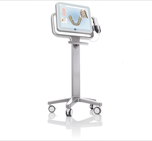
What Are iTero Intraoral Scanners?Intraoral scanners from iTero scan the mouths of patients, capturing images to create three-dimensional dental images in minutes. Intraoral scanners are simple to use and can be operated by one person. Their user-friendly nature helps dental professionals get the best results. The scans they produce are also more detailed than the traditional two-dimensional images they replace.Intraoral digital scans help dental professionals create accurate physical dental models for restorative work, including crowns, veneers, and implants. They also help orthodontists diagnose orthodontic problems and develop the best treatment plans.The company’s digital ecosystem software works seamlessly with its intraoral scanners, improving workflow for dental professionals working on orthodontic and restorative cases. However, unlike many early intraoral scanners, they are open systems. This feature gives dental professionals more flexibility about how they use their digital scan files. Intraoral digital scans from iTero scanners can be easily shared with other dental professionals and third-party providers such as Invisalign. When all relevant parties have the scans, they can communicate better to improve patient outcomes.What iTero Intraoral Scanners DoIntraoral scanners feature a wand, which the dental professional moves around a patient’s mouth. In the latest versions, the wand captures thousands of frames per second which are pieced together to create a three-dimensional visualisation of the patient’s mouth. The wands on iTero intraoral scanners are smaller than early intraoral scanners, allowing them to scan molars in the back of the mouth which were traditionally difficult to reach. Dental professionals using small wands also aren’t limited by how wide their patients can open their mouths. The small wands are also less likely to make patients gag than older forms of scanning technology.Intraoral scanners also have screens which display the digital dental images as they’re captured in real time. The screens show whether the scan is good or not before it’s saved and submitted to the lab. This feature can be a real time-saver for dental professionals who, in the past, could receive word from the lab two or three weeks later that their scans were inadequate. By providing immediate feedback, intraoral scanners can save dental professionals and patients time and frustration.Unlike many intraoral scanners, patients don’t need to cover their teeth in titanium dioxide powder before an iTero intraoral scan. This benefit further improves the scanning process for dental patients.How Intraoral Scanners Make Invisalign Orthodontics Easier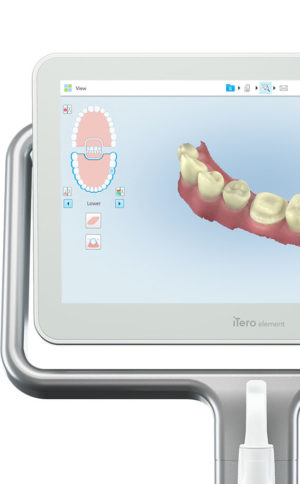 Invisalign clear aligners are one of the most popular teeth-straightening aids used today as they’re effective, removable, and virtually undetectable. Unlike many intraoral scanners, iTero intraoral scanners have open architecture which makes them compatible with the Invisalign system, including its Invisalign Outcome Simulator. Orthodontists can scan their patients’ mouths with an iTero intraoral scanner, then show them how their Invisalign treatment will look. This technology improves the patient experience because patients can know what to expect and feel more confident in their diagnosis and treatment plan. It also makes the ClinCheck setup three times faster.After setup, speed is still on the iTero intraoral scanners’ side. iTero states ClinCheck treatment plans submitted with its scans are usually posted to the Invisalign Doctor Site three times faster than traditional polyvinyl siloxane scans. As a result, your Invisalign aligners are created and posted back to your orthodontist sooner so that you can start treatment faster. Since the iTero intraoral scanning system is open, orthodontists can also send the scan files to any laboratory of their choosing for the creation of a retainer, which reinforces your treatment after the Invisalign process, and other dental tools.Orthodontists can also create better Invisalign treatment plans for their patients using iTero intraoral scans. Align Technology research shows orthodontists who use the scans have 10 times fewer rejections and seven times fewer issues with the fit of the Invisalign aligners. These results may be because iTero intraoral scanners can help orthodontists track their patients’ progress. Regular scans throughout Invisalign treatment can help orthodontists compare expected outcomes with results. If results aren’t as expected, orthodontists can use the scans to educate their patients about their treatment and the importance of complying with their recommendations.Improving iTero Intraoral Scanners iTero Element Intraoral ScannerWhile it’s already a leader in dental technology, iTero is constantly refining its intraoral scanning options. Its Element Itraoral Scanner, introduced in March 2015, captures 6,000 frames per second, up to 20 times faster than its predecessor. Its wand is also smaller and lighter, with in-built controls for more intuitive operation. Intraoral scanners from iTero are renowned for their accuracy, but the increased capture speed improves the company’s already impressive statistics. Invisalign clear aligners are one of the most popular teeth-straightening aids used today as they’re effective, removable, and virtually undetectable. Unlike many intraoral scanners, iTero intraoral scanners have open architecture which makes them compatible with the Invisalign system, including its Invisalign Outcome Simulator. Orthodontists can scan their patients’ mouths with an iTero intraoral scanner, then show them how their Invisalign treatment will look. This technology improves the patient experience because patients can know what to expect and feel more confident in their diagnosis and treatment plan. It also makes the ClinCheck setup three times faster.After setup, speed is still on the iTero intraoral scanners’ side. iTero states ClinCheck treatment plans submitted with its scans are usually posted to the Invisalign Doctor Site three times faster than traditional polyvinyl siloxane scans. As a result, your Invisalign aligners are created and posted back to your orthodontist sooner so that you can start treatment faster. Since the iTero intraoral scanning system is open, orthodontists can also send the scan files to any laboratory of their choosing for the creation of a retainer, which reinforces your treatment after the Invisalign process, and other dental tools.Orthodontists can also create better Invisalign treatment plans for their patients using iTero intraoral scans. Align Technology research shows orthodontists who use the scans have 10 times fewer rejections and seven times fewer issues with the fit of the Invisalign aligners. These results may be because iTero intraoral scanners can help orthodontists track their patients’ progress. Regular scans throughout Invisalign treatment can help orthodontists compare expected outcomes with results. If results aren’t as expected, orthodontists can use the scans to educate their patients about their treatment and the importance of complying with their recommendations.Improving iTero Intraoral Scanners iTero Element Intraoral ScannerWhile it’s already a leader in dental technology, iTero is constantly refining its intraoral scanning options. Its Element Itraoral Scanner, introduced in March 2015, captures 6,000 frames per second, up to 20 times faster than its predecessor. Its wand is also smaller and lighter, with in-built controls for more intuitive operation. Intraoral scanners from iTero are renowned for their accuracy, but the increased capture speed improves the company’s already impressive statistics. | Dexis Intraoral Digital Dental X-Ray SensorProduct DescriptionDexis Intraoral Digital Dental X-Ray SensorSoftware Ease of Use
According to the manufacturer, the DEXIS x-ray software was designed to be highly effective throughout diagnosis, treatment planning, and treatment. It is intended to serve as an imaging hub, managing all digital images, including intra- and extraoral radiographs and photographs. The manufacturer states that the images can be quickly organized, stored, retrieved, printed, and shared with patients and colleagues. The 9 dentists who participated in this product evaluation were asked to rate the DEXIS Digital Imaging software on its ease of use with regard to taking and accessing radiographs and for diagnosis, patient education, and communication with colleagues and insurance companies. Six of the evaluators rated the system as excellent and 3 rated it as very good. One evaluator commented that “when communicating with patients, we use DEXIS images displayed on a monitor in front of the patient every day on virtually every patient.” Software Integration
The manufacturer claims that the DEXIS imaging software can be easily integrated with all major practice management systems. According to DEXIS, the DEXIS Integrator automatically synchronizes with the patient record.When the evaluators were asked to rate the software integration with practice management software and interface with hardware (eg, cameras, panoramics, and scanners), 78% rated it as excellent to very good, while 22% rated it as good. One of the evaluators said, “The bridge to our practice management systems is seamless. We use [the] DEXIS digital pan[oramic] so that we can store panoramic x-rays easily in the same location as full series. We use an intraoral camera to store preoperative and interoperative photographs for excellent documentation.” Sensor Comfort and Ease of Use
According to DEXIS, the heart of their digital system is the PerfectSize sensor. The manufacturer states that their single-size sensor has 3 advantages:
1) because the electronics are on the back of the sensor, the active imaging area is most of the total sensor size;
2) the beveled corners, slim profile, and rounded casing allow for accurate placement and patient comfort;
3) patented cord placement allows the practitioner to take vertical and horizontal bitewings and all periapicals with only 1 universally sized sensor.
The evaluators were asked to rate both the ease of use of the sensor and patient comfort. Five evaluators rated ease of use excellent and 4 rated it very good. With regard to patient comfort, 3 evaluators rated the sensor excellent and 6 rated it very good. One evaluator noted that he experienced a few issues when placing the sensor in some children and patients with very small mouths. Another evaluator, however, is very happy with the sensor, saying, “I chose DEXIS to take a full radiographic series on every patient. It was the only direct image system we tested that met that objective.” Image Quality
According to DEXIS, their digital xray system provides detailed image quality and includes image enhancement tools that increase diagnostic capabilities. When the 9 evaluators were asked to rate the system’s image quality and enhancement aids, 89% rated them as excellent to very good. One of the evaluators commented that the system’s “ClearVu and image magnification allow us to diagnose very effectively.” Overall Satisfaction
When asked to rate their overall satisfaction with the DEXIS Digital X-ray System, 89% of the evaluators rated it as excellent to very good. Commented one evaluator, “DEXIS has been very responsive to our needs.” System Includes* a highend CCD sensor
* portable PC card and DEXusb connection options
* DEXIS imaging software
* a complete set of sensor holders and accessories
* on-site training
* a printed instruction manual
* training tutorials on CD
* a quick reference guide for the operatory
* 1 year of DEXcare support. | Schick CDR PanX Laser
FeaturesCDRPanX features proven Schick CDR digital imaging technology and renders crisp, clinically accurate images
Only slightly more expensive than most film-based systems, CDRPanX eliminates costly panoramic film and processing – increasing your economic efficiency
Eight different imaging modes and three laser alignment beams for optimal exposures
Superior vertical travel allows it to be used with patients of any height, including those in wheelchairs
Patient Positioning: 3 laser alignment beams (mid-sagittal, Frankfort, focal trough) bite guide, chin rest, optional temple support
SpecificationsGenerator: Multipulse DC
X-Ray Tube: OCX105
Focal spot size: 0.5 IEC 336
Minimum total filtration: 2.5 mm Al
Anode Voltage: 60-86kV (2kV increments)
Anode Current: 4, 5, 6.3, 8, and 10mA
Exposure time: 19s – adult panoramic
Power Supply: 115V, 50/60Hz
Fusing: T8A
Weight: 474lbs
Electrical Safety Classification: Class 1, type B
Imaging modes (Exposure times): adult (19s), child (15s), left half (10s), right half (10s), anterior (8s), TMJ opened and closed (4x4s), frontal sinuses (8s)
Patient Positioning: 3 laser alignment beams (mid-sagittal, Frankfort, focal trough) bite guide, chin rest, optional temple supportFeatures- CDRPanX features proven Schick CDR digital imaging technology and renders crisp, clinically accurate images
- Only slightly more expensive than most film-based systems, CDRPanX eliminates costly panoramic film and processing – increasing your economic efficiency
- Eight different imaging modes and three laser alignment beams for optimal exposures
- Superior vertical travel allows it to be used with patients of any height, including those in wheelchairs
- Patient Positioning: 3 laser alignment beams (mid-sagittal, Frankfort, focal trough) bite guide, chin rest, optional temple support
Specifications Schick CDR PanX Laser| Generator: | Multipulse DC | | X-Ray Tube: | OCX105 | | Focal spot size: | 0.5 IEC 336 | | Minimum total filtration: | 2.5 mm Al | | Anode Voltage: | 60-86kV (2kV increments) | | Anode Current: | 4, 5, 6.3, 8, and 10mA | | Exposure time: | 19s – adult panoramic | | Power Supply: | 115V, 50/60Hz | | Fusing: | T8A | | Weight: | 474lbs | | Electrical Safety Classification: | Class 1, type B | | Imaging modes (Exposure times): | adult (19s), child (15s), left half (10s), right half (10s), anterior (8s), TMJ opened and closed (4x4s), frontal sinuses (8s) | | Patient Positioning: | 3 laser alignment beams (mid-sagittal, Frankfort, focal trough) bite guide, chin rest, optional temple support |
|


Reviews
There are no reviews yet.