View cart “Energist NeoGen PSR Laser For Sale” has been added to your cart.
-41%
Availability: In Stock
PlanScan CAD/CAM Scanner Intraoral For Sale
$7,150.00$4,200.00
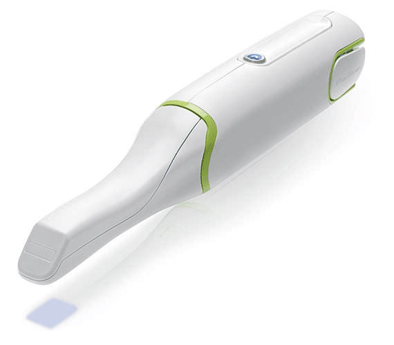
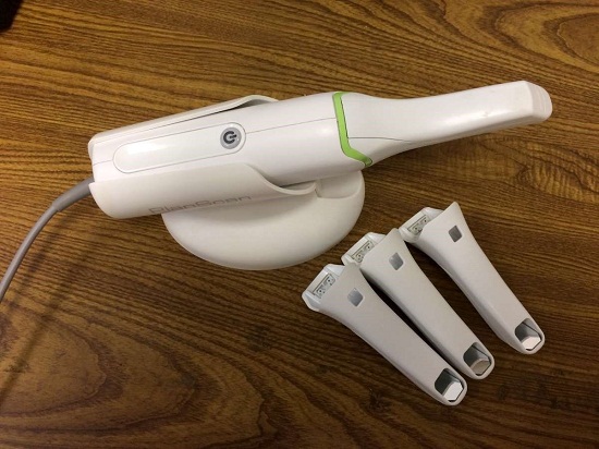
Explore Planmeca PlanScan – the first dental unit integrated intraoral scanner for digital 3D impressions. This high-performance intraoral scanning solution can also be connected to your laptop.
Other people want this. 15 people have this in their carts right now.
Excellent usability and workflow:
– Powder-free scanning
– Extremely easy to use
– Real-time, quick scanning
– True dental unit integration
– Autoclavable tip for perfect infection control
– Accurate results from single indication to full arch
– Comfortable solution
– Open STL format
– Windows support
– Works with a laptop – easy to share
Superior scanning speed
Planmeca Emerald is a blazingly fast intraoral scanner that allows full arches to be scanned in a few minutes or even less. The scanner’s active antifog feature ensures that Planmeca Emerald is always immediately ready to be used in seconds after plugging it in.
Ergonomic and lightweight design
Planmeca Emerald is exceptionally small and smooth to use. It has been designed to fit the user’s hand and places minimal strain on the wrist due to its light weight. The intraoral scanner’s small size is also very comfortable for patients.
True digital accuracy
Planmeca Emerald produces true-to-life colours with flawless detail. The scanner is superbly accurate and allows restorations and appliances designed onto digital impressions captured with it to fit flawlessly.
Two tip sizes PlanScan CAD/CAM Scanner Intraoral
In addition to the scanner’s ergonomic standard tip, Planmeca Emerald also has an even thinner tip available. The SlimLine tip is ideal for patients with smaller mouths and makes reaching posterior teeth and capturing interproximal areas easier. Both tips are autoclavable.
Flexible imports and exports
Planmeca intraoral scanners support imports and exports in STL file format, as well as PLY exports – making digital impressions captured with them easy to share with partners either in or outside a clinic without click fees.
For cooperation with dental labs, Romexis LabApp offers an easy and secure application for sending intraoral scans or other types of image data.
Comfortable scanning experience
Planmeca Emerald is comfortable for patients and users alike. The intraoral scanner’s small size and light weight support ergonomic chairside work and its speed ensures that scanning sessions remain short.
Various workflow possibilities
Our intraoral scanners offer unmatched flexibility. You can capture quadrants or full arches and either complete the entire restorative workflow on-site or conveniently export any part of it to your external partners.
Reviews (0)
Reviews
There are no reviews yet.
Be the first to review “PlanScan CAD/CAM Scanner Intraoral For Sale” Cancel reply
Explore Planmeca PlanScan – the first dental unit integrated intraoral scanner for digital 3D impressions. This high-performance intraoral scanning solution can also be connected to your laptop.
CIRRUS HD-OCT 5000 is configured with the highest resolution visualization capabilities and sophisticated clinical applications such as Advanced RPE analysis to track retinal pigment epithelial integrity and Ganglion Cell Analysis to assess glaucomatous loss in the macula that may not be evident in the peripapillary region. The new FastTrac retinal tracking reduces eye motion artifacts without sacrificing patient throughput with a proprietary scan acquisition strategy, high speed 20 Hz LSO camera, and single-pass alignment scanning. This feature enables the highest resolution B-Scans to be captured in identical locations from visit to visit providing precise assessment of change in targeted pathologies.
INCLUDES:
Picasso Lite+ Laser 3.0 Watts
Wireless Foot Control
Quick Start Guide
Owner’s Manual
Universal Power Supply
One Transportation Box
One Multi-Tip Handpiece
Three Pairs of Protective Laser Goggles
3 Year Warranty
The E4 boasts double the speed, double the accuracy, and double as many cameras as its predecessor the E3, delivering improved efficiency and consistency with every scan. From small labs seeking the best-of-the-best, to high-volume, full-service labs striving for a leaner workflow, our E4 lab scanner is the right choice for you.
Milling Unit:• Choose from CEREC MC, CEREC MC X or CEREC MC XL
• Get access to the entire range of chairside treatment with up to 40 mm block size, including bridges and abutments
• Create fully anatomic, precise treatments quickly
• CEREC Guide 2 surgical guide
• Dry mill ready
• Includes CEREC software
Ezer ERK-7800 Auto Refractor/KeratometerConfidently measure the human eye with a sophisticated cutting edge technology designed to enhance your ability to diagnose ocular errors faster, easier, and accurately. Ezers ERK-7800 is brilliantly designed technology measures the eye with ultra precision and speed to provide reliable data as the starting point of your refraction and contact lens fitting. It’s compact and modern design enables seamless operation and an enhanced patient comfort, making it the ideal choice in for any ophthalmic practice. Backed by an industry leading 2-years warranty!
Excellent usability and workflow:
– Powder-free scanning
– Extremely easy to use
– Real-time, quick scanning
– True dental unit integration
– Autoclavable tip for perfect infection control
– Accurate results from single indication to full arch
– Comfortable solution
– Open STL format
– Windows support
– Works with a laptop – easy to shareSuperior scanning speedPlanmeca Emerald is a blazingly fast intraoral scanner that allows full arches to be scanned in a few minutes or even less. The scanner’s active antifog feature ensures that Planmeca Emerald is always immediately ready to be used in seconds after plugging it in.Ergonomic and lightweight designPlanmeca Emerald is exceptionally small and smooth to use. It has been designed to fit the user’s hand and places minimal strain on the wrist due to its light weight. The intraoral scanner’s small size is also very comfortable for patients.True digital accuracyPlanmeca Emerald produces true-to-life colours with flawless detail. The scanner is superbly accurate and allows restorations and appliances designed onto digital impressions captured with it to fit flawlessly.Two tip sizes PlanScan CAD/CAM Scanner IntraoralIn addition to the scanner’s ergonomic standard tip, Planmeca Emerald also has an even thinner tip available. The SlimLine tip is ideal for patients with smaller mouths and makes reaching posterior teeth and capturing interproximal areas easier. Both tips are autoclavable.Flexible imports and exportsPlanmeca intraoral scanners support imports and exports in STL file format, as well as PLY exports – making digital impressions captured with them easy to share with partners either in or outside a clinic without click fees.
For cooperation with dental labs, Romexis LabApp offers an easy and secure application for sending intraoral scans or other types of image data.Comfortable scanning experiencePlanmeca Emerald is comfortable for patients and users alike. The intraoral scanner’s small size and light weight support ergonomic chairside work and its speed ensures that scanning sessions remain short.Various workflow possibilitiesOur intraoral scanners offer unmatched flexibility. You can capture quadrants or full arches and either complete the entire restorative workflow on-site or conveniently export any part of it to your external partners.
Carl Zeiss Cirrus HD-OCT 5000 Smart Advanced Topography HD
Advancing Smart OCT
for Anterior Segment, Glaucoma and Retina
Anterior Segment Premier Module
PanoMap Wide-Field Display
Smart HD Scans
En face report
Visualization at the speed of CIRRUS
Analyzing a single pathology from multiple views provides comprehensive insight and analysis of the clinical situation. How this helps you:
Spot small areas of pathology. Tightly spaced B-scans, (either 30 or 47 μm apart), in the cube ensure that small areas of pathology are imaged. For reference, a human hair is about 40-120 μm in diameter.
Visualize the fovea. Scans that are spaced further apart than in the CIRRUS cube may miss the central fovea.
Fuel for analysis. Millions of data points from the cube are fed into the Zeiss proprietary algorithms for accurate segmentation, reproducible measurements and registration for change analysis.
Take the pressure off the operator. As long as the scan is placed in the vicinity of the fovea or optic nerve, the software automatically centers the measurements after the capture.
See the tissue from different perspectives. View the cube data from all angles, with 3D rendering, OCT fundus images and Advanced Visualization™.
Future ready. Previously captured CIRRUS cubes can be analyzed using new analyses.
Tracking at the speed of CIRRUS
FastTrac™ reduces eye motion artifacts without sacrificing patient throughput with a proprietary scan acquisition strategy, high speed 20 Hz LSO camera, and single-pass alignment scanning
With FastTrac, scan at the highest resolution at the same location at each visit.
Watch video
Assessment at the speed of CIRRUS
Measurement centering with FoveaFinder and AutoCenter
On the macula, the unique FoveaFinder technology ensures the ETDRS and ganglion cell plus inner plexiform layer measurement frameworks are centered on the fovea.AutoCenter™ function automatically centers the 3.4 mm diameter peripapillary RNFL calculation circle around the disc for precise placement and repeatable registration. The placement of the circle is not operator dependent. Accuracy, registration and reproducibility are assured.
Manage at the speed of CIRRUS Carl Zeiss Cirrus HD-OCT 5000 Smart Advanced Topography HD
Guided Progression Analysis (GPA™) for Assessing RNFL and Optic Disc Change
GPA™ compares RNFL thickness and optic disc measurements over time and determines if statistically significant change has occurred.Focal Progression Analysis – Up to 6 progression maps are compared to two baselines. Areas of statistically significant change are color-coded yellow when first noted and then red when the change is sustained over consecutive visits.
Trend Analysis with Rate of Change – RNFL thickness values are plotted over time. Trends are calculated and confidence intervals are shaded when rate of change is statistically significant.
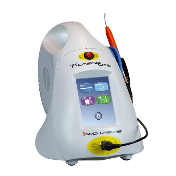
Feature & Benefits-3.0 Watts-Wireless Foot Control-Color Touch Screen-Easy to Use Presets-Built-In Treatment Timers for Periodontal Treatment and Aphthous Ulcer Treatment-Customizable Continuous/ Pulsed Modes-Multiple Languages Menu-Adjustable Volume & Aiming Beam Intensity-Two-Year Warranty*-Ideal for First-Time and Expert Laser Practitioners-Performs a Wide Range of Soft-Tissue Surgical Procedures & Periodontal Treatment-Offers the Convenience of Disposable Fiber Tips or Low-Cost Strippable Fiber*Warranty – 2 years parts and labor on the laser unit. Other terms and conditions apply.See the Owner’s Manual for complete details.Procedures -Gingival Troughing for Crown Impressions; Gingivectomy & Gingivoplasty-Gingival Incision & Excision; Hemostasis & Coagulation-Excisional & Incisional Biopsies-Soft-Tissue Crown Lengthening & Fibromal Removal-Frenectomy & Frenotomy-Implant Recovery & Exposure of Unerupted Teeth-Incision & Drainage of Abscess-Pulpotomy as an Adjunct to Root Canal Therapy & Reduction of Gingival Hypertrophy-Operculectomy & Vestibuloplasty-Leukoplakia & Oral Papillectomies-Treatment of Canker Sores, Herpetic & Aphthous Ulcers of the Oral Mucosa– Sulcular Debridement– Laser Soft-Tissue Curettage– Removal of Highly Inflamed Edematous Tissue Affected by Bacteria Penetration of the Pocket Lining & Junctional EpitheliumINCLUDES:
Picasso Lite+ Laser 3.0 Watts
Wireless Foot Control
Quick Start Guide
Owner’s Manual
Universal Power Supply
One Transportation Box
One Multi-Tip Handpiece
Three Pairs of Protective Laser Goggles
3 Year Warranty
THE MOST AFFORDABLE LASER IN THE DENTAL INDUSTRY
Keeping up with technology and implementing a dental laser into your practice sounds like a huge expense, but AMD Picasso Lasers are not only the most awarded soft-tissue lasers in the industry, but also the best priced--we guarantee! It’s no wonder Picasso Lasers are the most utilized soft-tissue lasers on the market.
3Shape E4 Dental Lab ScannerThe E4 is a dental 3D scanner produced by 3Shape. 3Shape is a 3D scanner manufacturer based in Denmark.E4 dental scanner main features
Full arch scan in 9 seconds
4-micron accuracy
Four 5-MP cameras
Color acquisition
3SHAPE E4
Ultimate productivity with our fastest scanner ever
For those seeking the speed and accuracy to drive lab productivity and growth, look no further. The E4 boasts double the speed, double the accuracy, and double as many cameras as its predecessor the E3, delivering improved efficiency and consistency with every scan. From small labs seeking the best-of-the-best, to high-volume, full-service labs striving for a leaner workflow, our E4 lab scanner is the right choice for you.
4 micron
Scan with an accuracy of 4 microns (ISO 12836) for great case results. Combine this precision with increased speed and you can take on more implant cases, for example.
4 cameras
Four cameras save you time by scanning dies in the model instead of individually. You can even scan impressions instead of producing a gypsum model or combine impression and die at the same time.
5MP cameras
5MP cameras are super high definition cameras. They record 5 megapixel images with enhanced camera sensors, meaning you get superior image clarity and can do high-precision scanning.
HARDWARE SPECIFICS
The E4 is a marriage of iconic 3Shape design and superior technical specifications.
Cameras
Four 5 MP cameras enables efficient die scanning and impression scanning, improving your workflows by saving time
Accuracy
4 microns (ISO 12836), giving you the accuracy and precision you need for implant cases
Full arch scan speed 9 seconds (versus 22 seconds for the 3Shape E3)
Full arch impressions scan speed 45 seconds
Texture Color
Scanning strategy Die-in-model
CEREC Primescan AC Intraoral scanner
Cerec Primescan from Dentsply Sirona is the most advanced digital scanner on the market. It’s providing a level of accuracy never before seen in dentistry. At the peak of innovation, it is widening the gap between those with and without digital dentistry. The Prime Scan is the fastest most accurate intraoral scanner to date. With new scanning technology, it is the ultimate in scanning. Prime Scan and CEREC 5.0 combinations are greatly reduced because more data allows many applications to become automated.For more than 30 years, CEREC® has provided technological precision, superior design and excellent performance to thousands of practices. Now, experience a brand-new fully integrated solution with CEREC Primescan AC. Acquire precise digital impressions with Primescan, design your proposal on the Primescan AC using the intelligent software, then produce precise, smooth restorations with your choice of milling unit. Find the next level of productivity and patient care – it’s all here.How Does CEREC Primescan Work?
CEREC Primescan is an alternative to traditional intraoral dental impressions, which use a putty-like substance to take physical impressions of your teeth. This method does not allow for digital assessment of your mouth, and some patients find it the process of physical impressions to be uncomfortable.CEREC Primescan uses a wand-like device, loaded with powerful imaging sensors which can capture more than 50,000 images of your mouth per second. These images are quickly stitched and compiled into a 3D image of your teeth, jaw, gums, and mouth, and displayed on a high-definition screen for immediate assessment by our doctors.Using this sensor, our doctors can image your entire mouth up to 20mm in depth, including gums, blood, and bone structures, quickly and accurately. The Primescan software (as compared to the Omnicam) creates high-resolution 3D models to assess your oral health and create the very best crowns, implants, removable prosthesis, restorations, and even 3D prints. With just a few quick sweeps of the CEREC Primescan device, we’ll be able to generate an in-depth, modifiable 3D image of your mouth in minutes.The Benefits of CEREC Primescan:
An excellent choice for outstanding results: Primescan is your perfect starting point in digital dentistry. No matter how you would like to design your workflows, Primescan is the enabler for efficient digital workflows – both chairside in your practice and with your preferred partners.Well designed and perfectly compatible: Along with the high-performance intraoral scanner comes the new Acquisition Center. Together, they form a highly effective and most comfortable system – meeting your needs with two individual software configurations:So, why is CEREC Primescan such a big deal? Here are just a few improvements to this sophisticated system:Shorter wait times – We can quickly image your mouth without waiting for dental impressions to set, allowing us to treat each patient more quickly and effectively. A full jaw impression only takes 2 minutes. This adds up to less time spent in the dental chair.Real-time image processing – Our dentists can quickly adjust and modify scanned images using filters and other tools, leading to more accurate diagnostics and better overall patient outcomes.Extremely versatile – CEREC Primescan can be used for almost any dental procedure. From implant dentistry to the creation of CEREC same-day crowns, the straightening of teeth, whitening, development of dentures and partial dentures.CEREC Primescan AC with CEREC Software Sirona Advantages:
Enables all kinds of treatment, from a single tooth to full arch
With Primescan intraoral scanning is now more accurate, faster and easier
Allows an efficient workflow, both chairside and with preferred partners
Along with the high-performance intraoral scanner comes the new Acquisition Center, together forming an effective and most comfortable system, meeting your needs with two individual software configurations: Primescan AC with Connect Software (supports the connection to established partners workflows) and CEREC Primescan AC with CEREC Software (ideal for cabinets)
Smart Pixel Sensor technology – allows superior precision and excellent detail reproduction, processes more than 1 million 3D points per second, producing photorealistic and highly accurate data
Due to its ability to scan at an incredible data density, it delivers complete 3D structures of everything in its field of view
Its dynamic depth scan technology enables perfect sharpness and outstanding precision, even at a measuring depth up to 20 mm
Easy to use
Easy scanning of all dental materials and hard to access areas
Allows you to start scanning right away
Self-heating for fog-free scanning
The increased field of view visualizes larger areas with fewer sweeps and immediate precision
Faster scanning with a smooth scan flow
Consolidating more than 50.000 images per second and fast processing of exactly the data the software needs Easy capturing and quicker processing of more data in higher resolution
Complete 3D-scan models are displayed immediately, no matter how fast you scan
Accelerate your workflow: Due to seamless, validated and open data transfer options, labs and other third parties are supplied with high-resolution models at an instant
3 different sleeves (disposable and stainless steel, with sapphire glass or autoclavable)
Acquisition Center – Smart features and greater comfort: intuitive use via movable 16:9 touchscreen and touchpad for perfect ergonomics
Kinematics for perfect ergonomic positioning
Smart Hygiene concept for fast and easy disinfection
Full mobility with more than 60-min. battery buffer
CEREC Primescan AC with CEREC Software – complete chairside system, which offers flexible data export options, with automatized workflow thanks to Artificial Intelligence and touch-enabled and intuitive user interface
Milling Unit:• Choose from CEREC MC, CEREC MC X or CEREC MC XL
• Get access to the entire range of chairside treatment with up to 40 mm block size, including bridges and abutments
• Create fully anatomic, precise treatments quickly
• CEREC Guide 2 surgical guide
• Dry mill ready
• Includes CEREC softwareStriking in every detail: The new Acquisition CenterThe new Acquisition Center is a work station designed for modern dentistry and with the dentist in mind. It comes with a touchpad and a 16:9 wide-format movable touchscreen, offering you a highly intuitive and ergonomic work platform.Your benefits at a glance:
Touchscreen & Touchpad for most comfortable, intuitive use
Kinematics for perfect ergonomic positioning
Smart Hygiene concept for fast and easy disinfection
Mobility concept for full mobility with more than 60-min. battery buffer
The best choice for your practice: Take digital impressions and continue working with your trusted partners or enjoy the benefits of complete chairside workflows.Primescan meets your needs with two individual software configurations:Primescan AC with Connect Software:
Supports data transfer options to your preferred partners
Secure and encrypted data transfer through Connect Case Center Inbox
Easy upgradability to full chairside workflow
Touch-enabled and intuitive user interface
CEREC Primescan AC with CEREC Software:
Supports full chairside workflows for single-visit dentistry
Flexible data export options
Automatized workflow thanks to Artifical Intelligence
Touch-enabled and intuitive user interface
Technical Data:CEREC Primescan AC Intraoral scanner
CAD system for high-precision intraoral optical impressions
High-resolution, heated intraoral 3D scanner
Integrated image processing
High processing power due to state-of-the-art processor
Trackball or touchpad
Hand and foot controlled enter keys
Ethernet port and WLAN
USB interfaces
The high-resolution 3D intraoral scanner allows image acquisition with the image control inside the scanner, the image data transfer is made through USB 2.0 interface
21.5″ inch TFT LED flat-screen display, HD resolution: 1920 x 1080 pixels
Mobile housing, with no water or air connection required
Voltage: 100 – 240 VAC/50-60Hz
Nominal current: 5.0 – 2.1 A
Type of protection against electric shock: Class I device
Waterproof: no
Degree of contamination: 2
Installation category: II
Operating mode: continuous
Dimensions W x H x D: 408 (537) x 1190 x 443mm
Total weight: 38kg
Weight without monitor and battery: 31kg
Weight without battery: 36kg
PC components:
Processor: Intel
RAM: 32GB RAM
Hard disks: 1x PCIe SSD + 1x SATA HDD
Network card: Ethernet 10/100/1000MBit/s
Operating system: Windows 10, 64 bit
Installation OS: pre-installed, from the factory
Transportation and storage conditions:
Temperature: -25°C – + 60°C
Relative humidity: 10% – 75%
Air pressure: 700hPa – 1060hPa
Operating conditions:
Temperature: 10°C – 35°C
Relative humidity: 30% – 85%, no condensation
Air pressure: 700hPa – 1060hPa
Operating altitude: up to 3000m
Delivery form:
CEREC Primescan AC with CEREC Software – 1 piece
Ezer ERK-7800 Autorefractor KeratometerFeatures of the ERK-7800 Auto Refractor/Keratometer Auto Refractor/Keratometer
Enhanced Retro-illumination Imaging
Accurate Measurement of Iris & Pupils Diameters
Precise Temperature Sensor Compensation
Enhanced Measuring Speed
Comfortable Motorized Chin Rest
Automatic High Speed Cutting Printer
Simple One Touch Lock Knob
20 Touch & Tilt Color Monitor Display
Sophisticated TFT LCD Graphic User Interface
Easy Network Capability
Backed by an Industry Leading 2-years Warranty!
Auto Refractor/Keratometer
The ERK-7800 instantly reports the patient’s assessed eye power by combining the Auto Refractor/Keratometer functions into this one sharp instrument, making measurement a snap. This determines the refractive and keratometric properties of the human eye to assist eye care professionals in the process of prescribing corrective eyeglasses and contact lenses. Keratometry and Refractometry (RK Mode) Enhanced Retro-illumination Imaging Ezer ERK-7800 Autorefractor Keratometer
With the Enhanced Retro-illumination Imaging capability in the ERK-7800, you can treat and see abnormal crystalline lenses, cataracts, and scratches of corneas helping you to determine how healthy the patients eyes are with better clarity. This allows for observation of the eye condition of the cataract or contact lens surface. The system saves up to 2 images of each eye. Accurate Measurement of Iris & Pupils Diameters
The corneal diameter and base curve of contact lenses can be measured with this one instrument. Therefore, measurements of eye and prescriptions for glasses and contact lenses can be made more efficiently. Measuring Cornea Size (SIZE MODE) Wide Dioptric Measurement Range
Because the ERK-7800 covers a wide measurement range from -25D to +22D, even an examinee with strong myopia can be measured.
Enhanced Measuring Speed
Specially designed image chips have succeeded to make REF measuring speed faster reducing the overall examination time and increases throughput for complete examinations. Comfortable Motorized Chin Rest
Simply by pressing up/down buttons, user can set the height of measuring point comfortably. With the cutting edge auto sensor mechanism, you can track down, measure and focus the eye with out ever making a mistake. Automatic Printer Paper Cut
The printer is designed to execute, print, & cut data automatically at high speed for immediate results. Simple One Touch Lock Knob
With just the touch of a single button, the main body can easily be locked to its base. 20 Degrees Tilted LCD Monitor
The LCD Monitor tilts up to 20 degrees; a design made for comfort making. Excellent ergonomics makes it possible to operate the ERK-7800 while standing or sitting, for added convenience. Sophisticated TFT LCD Graphic User Interface
Sharp images with easy to understand icons make this 6.4-inch color TFT LCD interface user-friendly and enjoyable to work with.





 An excellent choice for outstanding results: Primescan is your perfect starting point in digital dentistry. No matter how you would like to design your workflows, Primescan is the enabler for efficient digital workflows – both chairside in your practice and with your preferred partners.Well designed and perfectly compatible: Along with the high-performance intraoral scanner comes the new Acquisition Center. Together, they form a highly effective and most comfortable system – meeting your needs with two individual software configurations:So, why is CEREC Primescan such a big deal? Here are just a few improvements to this sophisticated system:Shorter wait times – We can quickly image your mouth without waiting for dental impressions to set, allowing us to treat each patient more quickly and effectively. A full jaw impression only takes 2 minutes. This adds up to less time spent in the dental chair.Real-time image processing – Our dentists can quickly adjust and modify scanned images using filters and other tools, leading to more accurate diagnostics and better overall patient outcomes.Extremely versatile – CEREC Primescan can be used for almost any dental procedure. From implant dentistry to the creation of CEREC same-day crowns, the straightening of teeth, whitening, development of dentures and partial dentures.CEREC Primescan AC with CEREC Software Sirona Advantages:
 The new Acquisition Center is a work station designed for modern dentistry and with the dentist in mind. It comes with a touchpad and a 16:9 wide-format movable touchscreen, offering you a highly intuitive and ergonomic work platform.Your benefits at a glance:
 Auto Refractor/Keratometer


Reviews
There are no reviews yet.