View cart “Samsung Medison SonoAce R3 Ultrasound Machine For Sale” has been added to your cart.
-46%
Availability: In Stock
Philips EPIQ 7 Ultrasound System For Sale
$40,000.00$21,530.00
Phillips EPIQ 7 is the most advanced ultrasound to ever hit the market. * Phillips powerful xMatrix transducers allows for the very best imaging in the business. *4D capacities allow for the most robust image quality ever made.
Other people want this. 15 people have this in their carts right now.
Philips EPIQ 7 Ultrasound features our strongest architecture ever applied to ultrasound imaging — touching all facets of acoustic acquisition and processing, letting you truly experience ultrasound’s development to a more definitive modality. Philips EPIQ 7 ultrasound is supported by our family of proprietary xMATRIX transducers and also our leading-edge Anatomical Intelligence.
The EPIQ 7 ultrasound process is encouraged by xMATRIX transducers, the many leading-edge, versatile ultrasound transducer technologies, and nSIGHT Imaging design permits for advances like the world’s first panoramic volume penis scan. NSIGHT Imaging also strengthens the power of PureWave, useful in imaging difficult patients.
Advanced introductions such as Live 3D imaging provides superior MVA evaluation and its utility on the EPIQ 7 program is simplified by incorporating all imaging modalities at the X5-1 transducer along with simplified measurement tools. Or, all the attributes on the iU22 and iE33 are magnified with all the Epiq 7.
The Philips EPIQ 7 ultrasound system is the most up-to-date in premium ultrasound, offering ease of use, workflow, ergonomics, and reliability with a vast array of sophisticated general imaging and cardiology Q-Apps to measure ultrasound image info. The EPIQ 7 ultrasound system is encouraged by xMATRIX transducers, the most leading-edge, flexible ultrasound transducer technologies, and nSIGHT Imaging design allows for progress such as the world’s first scenic volume penis scan. NSIGHT Imaging also strengthens the power of PureWave, helpful in imaging technically difficult patients.
With the XMATRIX, quantitative tools, and processing capacity, the Epiq 7 is Philips latest and greatest in 2015. With nSight and XMATRIX transducers, they’ve advanced the technology to carry out the world’s first panoramic organ scan.
Phillips EPIQ 7 is the most advanced ultrasound to ever hit the market. * Phillips powerful xMatrix transducers allows for the very best imaging in the business. *4D capacities allow for the most robust image quality ever made.
Highly Precise, Non-Surgical, Medical Procedure System for Professionals. Doublo HIFU’s Speed is Innovative Performance! Treatment time is dramatically reduced with Hironic’s High-capacity Technology and with Doublo HIFU System you can treat twice the amount of patients than beforeNew Standard of
MFU System
1.No. 1 MFU proven by the patents2.MFU proven by cumulative sales and clinical cases3.Fastest treatment by Two-way round shooting and Auto Shot mode
Function • Gravity Sensor: You can operate the ultrasound system either in transversal layout or vertical layout.• Grid for estimation: With grid on the image area, you can easily estimate the size of the target substance like follicle fertilized egg etc.
MedCorp is a trusted source for new and refurbished ultrasound systems with a combined 50 years experience in the industry and a detailed process for ensuring that each system they sell is as good as the day it was manufactured.
Ultraformer III’s 7 clip-in transducer cartridges project uniform ultrasound beams directly to multiple layers underneath the skin to promote tighter collagen formation, tissue contraction and reduction in the volume of adipocytes that form bulging areas of tissue.Patients notice an immediate benefit in improved skin tone and texture, with ongoing improvements over 3 to 6 months
Traditionally difficult areas such as eye lifting are made easier with Ultraformer III’s specialised cartridges. It is possible for patients to experience up to 6mm of eyebrow lift, depending on their particular situation.
Acuson Cypress cardiovascular system PLUS delivers advanced ergonomics and superior performance in a highly portable package. The Cypress System PLUS is a highly miniaturized, all digital, phased array echocardiography system that provides complete studies and outstanding images – even on patients who are difficult to image.
Content
Philips EPIQ 7 Ultrasound System
A new era in Premium UltrasoundPhilips EPIQ 7 Ultrasound features our strongest architecture ever applied to ultrasound imaging — touching all facets of acoustic acquisition and processing, letting you truly experience ultrasound’s development to a more definitive modality. Philips EPIQ 7 ultrasound is supported by our family of proprietary xMATRIX transducers and also our leading-edge Anatomical Intelligence.The EPIQ 7 ultrasound process is encouraged by xMATRIX transducers, the many leading-edge, versatile ultrasound transducer technologies, and nSIGHT Imaging design permits for advances like the world’s first panoramic volume penis scan. NSIGHT Imaging also strengthens the power of PureWave, useful in imaging difficult patients.
Advanced introductions such as Live 3D imaging provides superior MVA evaluation and its utility on the EPIQ 7 program is simplified by incorporating all imaging modalities at the X5-1 transducer along with simplified measurement tools. Or, all the attributes on the iU22 and iE33 are magnified with all the Epiq 7.The Philips EPIQ 7 ultrasound system is the most up-to-date in premium ultrasound, offering ease of use, workflow, ergonomics, and reliability with a vast array of sophisticated general imaging and cardiology Q-Apps to measure ultrasound image info. The EPIQ 7 ultrasound system is encouraged by xMATRIX transducers, the most leading-edge, flexible ultrasound transducer technologies, and nSIGHT Imaging design allows for progress such as the world’s first scenic volume penis scan. NSIGHT Imaging also strengthens the power of PureWave, helpful in imaging technically difficult patients.With the XMATRIX, quantitative tools, and processing capacity, the Epiq 7 is Philips latest and greatest in 2015. With nSight and XMATRIX transducers, they’ve advanced the technology to carry out the world’s first panoramic organ scan.Philips EPIQ 7 SpecificationsGeneral Imaging Q-Apps
This system is new, manufactured in 2017, it is in original packaging, and comes with a 12-month warranty.
DOUBLO GOLD HIFU SYSTEMWhy choose Doublo HIFU System?
HIFU (High-Intensity Focused Ultrasound System) is a highly precise medical procedure using high-intensity focused ultrasound to heat and destroy the pathogenic tissue rapidly. The earliest widespread use of HIFU was as a treatment for prostate cancer.HIFU hybrid system for Face & Body
Doublo Gold is the ultimate HIFU system for precise Face Lifting & Body Contouring. Offering the fastest treatment by Two-way round shooting and Auto Shot mode.Non Invasive – No Pain – No Bleeding – No Downtime
Faster shot speed : Efficacy improved by repetitious shooting with 300 shots in 8 minutes. Entertain more patients with a faster treatment procedure.Multiple treatment areas
By treating the upper dermis with wound healing effect, facial pores and skin tone is improved as the fibroblast is rebuilt and collagen is boosted.Dermal tissue elasticity and collagen increase as fibroblast is rebuilt by wound healing effect from damaging sub-cutaneous tissue and derma layer with thermal coagulation.
Skin tightening to reduce wrinkle and increase lifting effect is done by creating thermal coagulation at subcutaneous fat and SMASDOUBLO GOLD HIFU SYSTEM Highly Precise, Non-Surgical, Medical Procedure System for Professionals. Doublo HIFU’s Speed is Innovative Performance! Treatment time is dramatically reduced with Hironic’s High-capacity Technology and with Doublo HIFU System you can treat twice the amount of patients than beforeDOUBLO GOLD HIFU SYSTEM Accessories: Power cord for cart
Clear plastic tray insert
Large washer and small lock/spring washer
2x black attachements
2x treatment parameters guide
Simple user manual
Full user manual + CDHighly Precise, Non-Surgical, Medical Procedure System for Professionals. Doublo HIFU’s Speed is Innovative Performance! Treatment time is dramatically reduced with Hironic’s High-capacity Technology and with Doublo HIFU System you can treat twice the amount of patients than beforeNew Standard of
MFU System
1.No. 1 MFU proven by the patents2.MFU proven by cumulative sales and clinical cases3.Fastest treatment by Two-way round shooting and Auto Shot mode
SIUI CTS-800 Veterinary Ultrasound Machine with Linear Rectal Probe
This small and convenient “Palm-Sized” portable veterinary ultrasound scanner fits in the palm of your hand, and is mostly used for farm animals. Its long battery life and waterproof features makes it the perfect ultrasound scanner for veterinarians on the field.The screen of the SIUI CTS-800V will rotate horizontally or vertically, according to how you are holding it.Gravity Sensor: You can operate the ultrasound system either in transversal layout or vertical layout. It can automatically adapt to two different layouts when you rotate it.Grid for estimation: With grid on the image area you can easily estimate the size of the target substance like follicle fertilized egg etc.SPECIFICATIONS SIUI CTS-800 Veterinary Ultrasound Machine with Linear Rectal ProbeMonitor: 7-inch WVGA LCD monitor
– Main unit with 7-inch LCD monitor.
– 1 Probe connector.
– 1 Li-ion battery. 3 hours operation time.
– 1 Adaptor.
– 1 Car Charger.
– Belt and sun shield.
– 1 USB port (Mini).
– Carrying case.PROBES– Rectal Linear L7FVC (6.5/7.1/7.5/8.3/9.0MHz).
– Rectal Linear L5FVC (4.0/4.7/5.0/5.5/6.2MHz).
– Backfat Linear L1FC (150mm, 2.5/3.0/3.5/4.0/5.0MHz).
– Convex C3FC (2.5/3.0/3.5/4.0/5.0MHz).
– Microconvex C5FC (3.5/4.0/5.0/6.5/7.1MHz).CTS-800 is a palm-sized veterinary ultrasound system. Features Weight: 0.8kg Environmental rating: IP54 (main unit) IP67 (probe head) Battery: 3 hours operation time Display mode: B 2B ZOOM B B/M M Depth: Max to 250mm B gain: 0-100 Image storage: 256 frames Cine loop: 256 frames Probe: L50/7.5 MHz linear rectal probe (standard) L65/5.0 MHz linear rectal probe (optional) R20/5.0 MHz micro convex probe (optional)
SonoSite Nanomaxx Ultrasound Machine
The SonoSite NanoMaxx ultrasound system combines premium functionality and affordability with the versatility and portability that makes SonoSite a top competitor in the portable ultrasound market. The SonoSite NanoMaxx comes in a sleek, hand-held package that never compromises performance, with applications ranging from emergency medicine and anesthesia to vascular.MedCorp offers the refurbished SonoSite NanoMaxx for purchase. Used SonoSite NanoMaxx from MedCorp offer everything you’d expect from a newer machine, including PC/MAC export configurations, 2D, Color and Color Power Doppler modes, and 2GB of internal memory that can hold 50 images each for 40 patients, all in an 8” touch-screen interface. However, because this is a refurbished SonoSite NanoMaxx, the price is lower than the new models.
Comes with:* NanoMaxx Ultrasound System, 1.0.1
* Battery – NanoMaxx 11.25 volt, 5.2 amp-hour rechargeable lithium-ion battery
* L38N / 10-5 MHZ Transducer multi-frequency, broadband, 38 mm linear array transducer for small parts, breast, vascular, nerve, IMT, and venous. Supports 2D with SonoMB Multi Beam Technology and Auto Gain capabilities, SonoHD Imaging Technology, Color Power Doppler, velocity Color Flow Doppler. Maximum depth is 9 cm.
* Power Supply, Universal supply (50-60 Hz, 100-240 VAC) for charging battery and powering the NanoMaxx.
* NanoMaxx Carry Case
8″ Touch-screen interface
2D, Color and Color Power Doppler modes
2GB internal memory that can hold 50 images each for 40 patients
2 hours of battery power
PC/MAC export configuration
Four preset pictograph screen positions
Drop resistant magnesium case
System Description:SonoSite Nanomaxx Ultrasound Machine
The NanoMaxx ultrasound system offers excellent performance at an affordable price. It is easy to use and offers top of the line diagnostic imaging, color power Doppler and color-flow velocity. The NanoMaxx can aid users in making clinical decisions or guide interventional procedures. The unit uses proprietary technology which allows the optimization of many system settings by simply pressing a button.Application:
Abdominal, Cardiology, Gynecology, Nerve, Obstetrics, CIMT, Musculoskeletal, Small Parts, Superficial, Vascular, Venous
You’ve heard about it, now see and feel the difference for yourself.Ultraformer III is the leading technology for facial lifting, tightening and contouring. Now with 7 interchangeable cartridges that offer your patients another dimension in non-surgical face and body treatments.Ultraformer III is widely used with over 10 million treatments performed worldwide. It has become the treatment of choice for patients who are not yet indicated or not willing to undergo surgery. This fills a huge gap in the market, and captures an audience of people from 30 years of age all the way to 80.ULTRAFORMER III FEATURESLess discomfort associated with treatments
Patented transducer design for more efficient energy delivery
Dual hand pieces with automatic recognition for ease of use
Faster shot and treatment speed to save time
Easy, user-friendly design and interface
Shot count information and voice assistance
Backed by clinical papersThe CLASSYS ULTRAFORMER III UTR3 advantage with slim transducer designimproves operator visibility
supports very narrow focus and precise treatment
fits facial contoursFast elliptic transducersmore efficient treatment timesUltraformer III’s 7 clip-in transducer cartridges project uniform ultrasound beams directly to multiple layers underneath the skin to promote tighter collagen formation, tissue contraction and reduction in the volume of adipocytes that form bulging areas of tissue.Patients notice an immediate benefit in improved skin tone and texture, with ongoing improvements over 3 to 6 months
Traditionally difficult areas such as eye lifting are made easier with Ultraformer III’s specialised cartridges. It is possible for patients to experience up to 6mm of eyebrow lift, depending on their particular situation.Ultraformer III’s 7 clip-in transducer cartridges project uniform ultrasound beams directly to multiple layers underneath the skin to promote tighter collagen formation, tissue contraction and reduction in the volume of adipocytes that form bulging areas of tissue.Patients notice an immediate benefit in improved skin tone and texture, with ongoing improvements over 3 to 6 months
Traditionally difficult areas such as eye lifting are made easier with Ultraformer III’s specialised cartridges. It is possible for patients to experience up to 6mm of eyebrow lift, depending on their particular situation.
Acuson Cypress PLUS Ultrasound
Acuson Cypress cardiovascular system PLUS delivers advanced ergonomics and superior performance in a highly portable package. The Cypress System PLUS is a highly miniaturized, all digital, phased array echocardiography system that provides complete studies and outstanding images – even on patients who are difficult to image.The Cypress PLUS transitions easily from your small private office or hospital echo lab to the operating theatre or your patient’s bedside. It’s ultrasound where you need it.TheCypress PLUS delivers high performance in a complete range of cardiovascular capabilities.Designed with advanced ergonomics in mind, the Cypress system PLUS streamlines your workflow and increases your productivity.If you already own a Cypress system, keeping technologically current is simple with the Cypress Upgrade program.The Cypress system is the smallest, lightest, high performance, phased array echo system ever built. Weighing in at just 19 pounds, with digital storage capacity for over 100 studies and network capabilities to send studies from remote locations, clinicians can perform complete echocardiography studies from almost anywhere. The Cypress system was designed for portability, durability and reliability, and to withstand the rigors of constant transportation. Internal system interconnects are minimized. Key components are shock mounted and the entire system sits inside a robust chassis. From hospital to office or from airplane to mountain village, the Cypress system will perform.Full Range of Capabilities
TheCypress system is a complete, full-featured echo cardiography system that provides complete studies and outstanding image quality even on the most difficult to image patients. It offers a full range of features including:– 2D with Harmonics
– Patient Report with audio capture
– M-Mode
– Embedded DICOM conformance
– Color PW and CW Doppler
– High frame rate color flow imaging
– Stress echo
– Network output
– Vascular imaging
– Comprehensive calculations package
– Contrast agent imaging
– USB peripheral support (PC printer connectivity)Physical Characteristics Acuson Cypress PLUS Ultrasound• Single unit houses ultrasound system with control panel, flat panel display and MO drive
• Weight: 8.6 Kg (19 pounds)
• Height: 35.6 cm (14 in)
• Width: 40.6 cm (16 in)
• Depth: 20.3 cm (8 in) – with keyboard folded up 34.3 cm (13.5 in) – with keyboard down
• Heat dissipation: 490 BTU/hour (nominal)User Interface
• Fold-up control panel for portability
• Direct access to system functions through dedicated keyboard controls and multifunctional soft keys
• Easy-access main controls, including multifunctional trackball, enter and select keys, for minimum hand movement while controlling the system
• Adjustable trackball speed
• Functional grouping of keys, rotational knobs, switches and sliding TGC controls for direct access to critical functions
• Tactile feedback on control activation
• Backlighting of control panel labels
• Mode-dependent soft keys
• User-interface tools
• Pre-defined and user-entered labeling textMonitor
• 10.4 inch/26.4 cm flat panel active matrix display screen
• Protective anti-glare, anti-reflective lensHard Drive
• Internal hard drive for image and data storage
• Internal storage of approximately 50 patient studies (25 – 30 loops per study)Transducer Ports
• One transducer connector (type IEC601BF)Operating/Display Modes
• 2-D, fundamental and harmonic imaging
• Color Doppler Velocity (CDV)
• Color Doppler Energy (CDE)
• M-mode, vertical split screen display with 2-D
• Pulsed Wave (PW) Spectral Doppler
• Continuous Wave (CW) Spectral Doppler
• Duplex Doppler (combined 2-D and Spectral Doppler display)2D Calipers – Generic Measurements and Calculations
• Measurements and calculations in 2D, M-mode, color Doppler, PW and CW spectral Doppler modes
• Basic measurements and report calculations
• Comprehensive patient calculations report
• On-screen “mini-reports”
• Complete or configured patient report
• Cardiac package
• Carotid package
• Arterial package
• Venous package
• Abdominal package
• User configurable Sites
• Selectable menu position
• Four cursor types
• Angle correction
• Distance
• Area
• Slope
• Time
• Velocity
• Velocity/pressure
• Mean velocity/pressure gradient
• Velocity Time Integral
• Pressure halftime
• Continuity Equation
• Ejection Fraction
• Left Ventricular Volumes, including End-Systolic and End-Diastolic volumes
• Cardiac output
• Cardiac index
• Fractional area changeCine Review
• 2D Cine memory up to 15 seconds of history
• Color Doppler cine memory up to 15 seconds of history
• M-mode strip cine along with PW and CW Doppler mode
• strip cine memory determined by sweep speed (selectable by the user) 10, 20, 30 or 40 secondsTransducer Technology
• 3V2c: 2-4 MHz wideband phased array
• 7V3c: 3-7 MHz wideband phased array
• 7L3: 5-7 MHz wideband linear array (Vascular)
• 4C1: 2-4 MHz wideband curved linear array (Abdominal)
• V5Ms: 3-6 MHz wideband phased array (micro-pinless (MP) connector)


 The Philips EPIQ 7 ultrasound system is the most up-to-date in premium ultrasound, offering ease of use, workflow, ergonomics, and reliability with a vast array of sophisticated general imaging and cardiology Q-Apps to measure ultrasound image info. The EPIQ 7 ultrasound system is encouraged by xMATRIX transducers, the most leading-edge, flexible ultrasound transducer technologies, and nSIGHT Imaging design allows for progress such as the world’s first scenic volume penis scan. NSIGHT Imaging also strengthens the power of PureWave, helpful in imaging technically difficult patients.
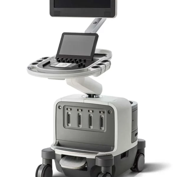


Reviews
There are no reviews yet.