| Content | Philips Affiniti 70 Ultrasound system
Philips Affiniti 70 ultrasound system for women’s healthcarePhilips Affiniti 70 Ultrasound system delivers a powerful mix of performance and workflow for diagnosis. Engineered for efficiency and dependability and powered by Philips demonstrated performance, it gets users diagnostic pictures, quickly on the most technically difficult patients. Its layout and walk-up usability provide efficient maintenance every day.The Philips Affiniti 70 is a complete echocardiography alternative that addresses the needs of a busy office or department while incorporating those inventions that make Philips ultrasound the international pioneer in echocardiography.Philips Affiniti 70 precision beamforming, PureWave technology, Tissue-Specific Presets (TSP), and efficiency and automation tools provide both functionality and workflow for confident throughput. Designed around your workflow, the Philips Affiniti 70 provides mobility, ergonomics, and usability.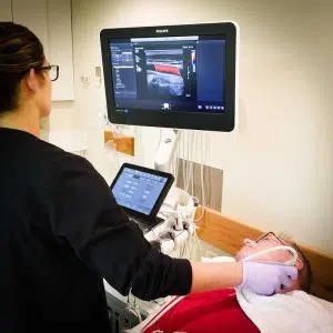 The Affiniti 70 also has many of the same technology as the Epiq series, including the amazing QLAB onboard quantification set and ShareWave elastography. The Philips Affiniti 70 is exceptional in the high-end price category in offering ShearWave. As a service ultrasound device that is shared the 70 excels at cardiology in addition to women’s health, vascular and radiology programs. Philips is well known for its imaging superiority over other brands, but the Philips Affiniti 70 is also rich with excellent image quality at its price level. Doctors who like the Affiniti 70, but do not need the transducers or ShearWave Elastography should consider the less costly Affiniti 50.The Philips Affiniti 70 combines outstanding performance and effective workflow and attributes innovative Philips technologies. The Precision Beamforming delivers outstanding tissue uniformity and contrast resolution, excellent spatial, few artifacts, and decreased image jumble. Tissue-Specific Presets (TSPs) mechanically adjust over 7500 parameters to optimize the transducer to your particular test type, making excellent picture quality with little if any demand for picture adjustment. The Affiniti 70 Machine can also be equipped with automation features, for example, AutoSCAN and automobile Doppler, which reduce repetitive tasks and make obtaining a picture. Exam efficiency is delivered by Philips SmartExam-guided workflow.Key Features: The Affiniti 70 also has many of the same technology as the Epiq series, including the amazing QLAB onboard quantification set and ShareWave elastography. The Philips Affiniti 70 is exceptional in the high-end price category in offering ShearWave. As a service ultrasound device that is shared the 70 excels at cardiology in addition to women’s health, vascular and radiology programs. Philips is well known for its imaging superiority over other brands, but the Philips Affiniti 70 is also rich with excellent image quality at its price level. Doctors who like the Affiniti 70, but do not need the transducers or ShearWave Elastography should consider the less costly Affiniti 50.The Philips Affiniti 70 combines outstanding performance and effective workflow and attributes innovative Philips technologies. The Precision Beamforming delivers outstanding tissue uniformity and contrast resolution, excellent spatial, few artifacts, and decreased image jumble. Tissue-Specific Presets (TSPs) mechanically adjust over 7500 parameters to optimize the transducer to your particular test type, making excellent picture quality with little if any demand for picture adjustment. The Affiniti 70 Machine can also be equipped with automation features, for example, AutoSCAN and automobile Doppler, which reduce repetitive tasks and make obtaining a picture. Exam efficiency is delivered by Philips SmartExam-guided workflow.Key Features:- 21.5” wide flat panel LCD display monitor
- Fully articulating arm
- 2” Tablet-like widescreen touchscreen
- 4 probe ports
- TDI [Tissue Doppler Imaging]
- THI [Tissue Harmonic Imaging]
- TAC [Tissue aberration correction]
- LVO [Left ventricular opacification]
- CPA and Directional CPA [Color Power Angio]
- Coded beamforming
- Steerable CW Doppler
- High-PRF PW Doppler
- Auto Doppler optimization
| Siemens / Acuson Sequoia 512 Ultrasound Machine
The ACUSON Sequoia 512 ultrasound is an ultra-premium system that utilizes revolutionary technology, dedicated to the advancement of diagnostic imaging. It represents some of the most advanced technology in ultrasound imaging. The Acuson Sequoia 512 has unparalleled imaging performance, greater image content, enhanced detail, superior contrast resolution & penetration, and excellent color Doppler sensitivity.One of the best selling ultrasounds in the United States, the Sequois 512 provides superior imaging with Native Patient Specific Imaging that detects, measures and adapts (in real time) to each patient’s individual acoustics.Performance:Reduce image optimization time by up to 80% and allow for an easier, faster and more confident diagnosis:- 2D
- SST Color Doppler
- Color Doppler Energy
- Color Doppler Velocity
- Solo Spectral Doppler
- M-mode and Color Doppler M-mode
- MultiHertz Multiple Frequency Imaging
- DELTA Differential Echo Amplification
- Home Base user interface
- One-Touch Image Control with exam and Image Presets
- Digital acquistion, storage, review and transfer of static images, dynamic clips, and cine
- Digital storage on an integrated hard disk
- Digital transfer via MO Disk
- Integrated Compression engine
- Embedded DICOM conformance
- Patient- oriented file structure
- Native Tissue Harmonic Imaging
- Tissue Equalization™ Technology (Optional)
- Signature II™ (Optional)
- Advanced Imaging package (Optional)
- Auto Doppler (Optional)
- Freestyle Compounding (Optional)
- FreeStyle Extended Imaging (Optional)
- 3D surface Rendering (Optional)
- Cadence™ ADI (Optional)
- Cadence CPS for 512 (Optional)
- Axius™ Edge Assisted EF (Optional)
- Doppler Tissue Imaging (Optional)
- DICOM Print And Store (Optional)
Capabilities:Siemens / Acuson Sequoia 512 Ultrasound MachineGeneral Radiology, Abdominal, Intraoperative Monitoring, OB/GYN, Neonatal, Vascular, Cardiac, Small PartsAvailable Transducers:4V1, 4V2, 8V5, 15L8W, 6L3, 8L5, 6C2, 5C2, 4C1, 3V2C, 5V2C,7V3C, EVC8-4V, EC-10C5, V5M TEE, V7B neo TEE, AcuNav Transducer, 2.0 Non Imaging CW Doppler probe.Acuson sequoia 512 ultrasound click images to enlarge item description acuson sequoia 512 ultrasoundsold as picturedincludes: 2 probes, 4c1 convex & 6l4 linearsony up 895 md printerunit will be cleaned, function checked and certified for electrical safety. Checked, cleaned, certified for electrical safetyguaranteed working when arrives on the guarantee working we need to be notified in 48 hrs. And the seals must be intact. | Samsung Medison SonoAce R3 Ultrasound Machine
SonoAce R3 is a portable color ultrasound system with comprehensive diagnostic capabilities. Full Spectrum Imaging™ (FSI™) combines with Speckle Reduction Filter™ (SRF™) to deliver superior quality images, even in the challenging circumstances. The compact and user-friendly design features of the SonoAce R3 ensure portability and manoeuvrability in a lightweight unit. Sporting a 15-inch LCD monitor with Full-Featured Workflow, the SonoAceR3 offers your organisation flexibility, without compromising on precision and efficiency.Slim and Compact DesignThe compact design of SonoAce R3 offers Various-Featured Workflow with a thumbnail menu and shortcut, together with functional key groupings for increased efficiency. The user-friendly design combines a wide-view 15-inch LCD monitor with backlit control keys, customisable measurement package and body markers. The SonoAce R3 features 2 probe ports, 3 USB ports, rapid boot up capability and DICOM 3 compatible image filing. This slim, lightweight unit also has a convenient carrying handle for portability and flexibility.Samsung Medison SonoAce R3 Ultrasound MachineFull Spectrum Imaging™ (FSI™) produces the penetration capabilities associated with the lower frequencies, while still maintaining the fine pixel uniformity associated with the higher frequencies. This combination consistently delivers superior quality images, even in the challenging diagnostic circumstances. The resulting clarity delivers improvements in accuracy and efficiency for greater confidence in your diagnostic procedures. Harmonic Imaging is a technique that records higher frequency information, resulting in greater resolution and fewer artifacts.Synthetic Aperture
The physical radio channel may present some limits to imaging which can be overcome by using Synthetic Aperture Control software. This method uses information from diverse sources to form a more better picture of the area under examination. It creates one single scan line by performing the TX and RX twice to create enhanced penetration and resolution. Synthetic Aperture Control technology delivers a comprehensive picture and to aid accurate diagnosis.QuickScan
QuickScan has been designed to increase workflow efficiency by automatically optimising key imaging parameters at the touch of a button. The resulting increase in efficiency allows your organisation to offer patients an improved service with faster and more accurate diagnoses. This reliable and safe system helps your clinic or hospital to deliver a superior patient experience.Features
Full Spectrum ImagingTM
Quick ScanTM
Color and Power Doppler
Harmonic imaging
Freehand 3D
3D Multi Planar Imaging
Sonoview Image Management
PW Doppler
15″ LCD monitor
Two probe ports
Lightweight | GE Voluson 730 Expert Ultrasound MachineGE Voluson 730 Expert, 3D/4D Realtime Ultrasound System Color Doppler, PW Doppler, Tissue Harmonics, STIC, VOCAL DICOM, M-mode, Angio, ATO, TUI, SonoView II, Crossbeam (CRI) MO Drive, Cineloop, High Resoluotion Interlaced Monitor Probe Options: RAB4-8 4D Convex RIC5-9 4D Transvaginal AB2-7 2D Convex IC5-9 2D Transvaginal SP6-12 2D Linear PA2-5 Cardiac Sector
Two Probes included:- RAB4-8L – Live 4D Abdominal Transducer
- RIC5-9 – Live 4D Transvaginal Transducer
Options:
About the Voluson 730:GE Voluson 730 Expert Ultrasound MachineWith GE Voluson 730, you can now acquire and construct volumetric images in real time – up to 40 volumes per second. The system allows you to explore images in any plane to reveal the smallest details with stunning clarity and apply sophisticated analytical tools to answer virtually any of your clinical questions. Volume Ultrasound not only improves your 3D/4D capabilities, it enhances your 2D imaging as well, for even greater diagnostic confidence in your obstetrical, gynecologic, breast and general imaging studies.The used GE Voluson 730 Expert was among the top-rated and biggest breakthrough ultrasounds in the industry. With up to 40 volumes per second rate of acquisition and construction of volumetric images in real time. The 730 Expert is loaded with the newest ultrasound technology including: CrossXBeam, Speckle Reduction Imaging, Tomographic Ultrasound Imaging (TUI), Spatio-Temporal Image Correlation (STIC), and much more.The Voluson 730 Expert is the more advanced version of the Voluson 730 Pro and was replaced by the GE Voluson e8. Compared to the 730 Pro, the Voluson 730 Expert features a touchscreen interface, higher frame rates in 2D and 4D, and more advanced imaging options such as STIC and TUI.Providian Medical Voluson 730 Expert Review:The Voluson 730 is a workhorse of a 4D ultrasound machine. It remains a popular system in many OB/GYN/Women’s Health offices, as well as 4D “Babyface” studios around the world. It is a durable machine with very good image quality for all women’s health needs. | Samsung Medison SonoAce R3 Ultrasound MachineSonoAce R3 is a portable color ultrasound system with comprehensive diagnostic capabilities. Full Spectrum Imaging (FSI) combines with Speckle Reduction Filter (SRF) to deliver superior quality images, even in the challenging circumstances. The compact and user-friendly design features of the SonoAce R3 ensure portability and manoeuvrability in a lightweight unit. Sporting a 15-inch LCD monitor with Full-Featured Workflow, the SonoAceR3 offers your organisation flexibility, without compromising on precision and efficiency.Slim and Compact DesignThe compact design of SonoAce R3 offers Various-Featured Workflow with a thumbnail menu and shortcut, together with functional key groupings for increased efficiency. The user-friendly design combines a wide-view 15-inch LCD monitor with backlit control keys, customisable measurement package and body markers. The SonoAce R3 features 2 probe ports, 3 USB ports, rapid boot up capability and DICOM 3 compatible image filing. This slim, lightweight unit also has a convenient carrying handle for portability and flexibility.Full Spectrum Imaging (FSI) produces the penetration capabilities associated with the lower frequencies, while still maintaining the fine pixel uniformity associated with the higher frequencies. This combination consistently delivers superior quality images, even in the challenging diagnostic circumstances. The resulting clarity delivers improvements in accuracy and efficiency for greater confidence in your diagnostic procedures. Harmonic Imaging is a technique that records higher frequency information, resulting in greater resolution and fewer artifacts.Synthetic Aperture
The physical radio channel may present some limits to imaging which can be overcome by using Synthetic Aperture Control software. This method uses information from diverse sources to form a more better picture of the area under examination. It creates one single scan line by performing the TX and RX twice to create enhanced penetration and resolution. Synthetic Aperture Control technology delivers a comprehensive picture and to aid accurate diagnosis.QuickScan
QuickScan has been designed to increase workflow efficiency by automatically optimising key imaging parameters at the touch of a button. The resulting increase in efficiency allows your organisation to offer patients an improved service with faster and more accurate diagnoses. This reliable and safe system helps your clinic or hospital to deliver a superior patient experience.Features Samsung Medison SonoAce R3 Ultrasound Machine
Full Spectrum ImagingTM
Quick ScanTM
Color and Power Doppler
Harmonic imaging
Freehand 3D
3D Multi Planar Imaging
Sonoview Image Management
PW Doppler
15″ LCD monitor
Two probe ports
Lightweight | Medison Accuvix XG Ultrasound System
Samsung Accuvix XG Ultrasound Machine2D Image Features
• Dynamic MR™ / Dynamic MR Plus is designed to enrich gray-scale resolution, as it enhances detection
and contrast resolution while also decreasing speckle echoes. This is particularly useful when evaluating
superficial structures, including thyroid, vessels, pelvic and abdominal anatomy.
• SRF (Speckle Reduction Filter™) enhances image quality by reducing or eliminating the appearance of
speckle echoes from ultrasound images. The degree of speckle reduction implemented is user-selectable.
• Wide Dynamic Range determines the number of gray shades utilized to map the gray-scale image.
It enables display of more details of bright areas and dark areas.
• SCI (Spatial Compounding Image) controls the ultrasound beam electronically by steering, and compounds
many scan lines.
• FSI (Full Spectrum Imaging) incorporates the penetration capabilities associated with lower frequencies,
yet maintains the fine pixel uniformity associated with higher frequencies, to deliver consistently high quality
images even in challenging diagnostic areas.3D Image Features Medison Accuvix XG Ultrasound System
• HDVI gives outstanding image quality and naturally clearer contrast, with excellent tissue differentiation,
edge depiction and speckle reduction, allowing consistent diagnoses with great confidence.
• 3D XI™, comprised of a suite of three innovative imaging applications – Multi-Slice View, Oblique View™ and
Volume CT – offers complete and precise control over 3D/4D volume data manipulation for maximum
diagnostic accuracy.
• 3D MXI is an innovative, cutting-edge 3D image processing technology. Comprising a comprehensive suite of
imaging tools – including Multi Volume Slice, Mirror View, Multi-OVIX, and 3D OH – 3D MXI lets you view,
examine and diagnose 3D volume data with supreme ease, speed and accuracy. • Fully adjustable system
The control panel can be adjusted to the user’s preferred height, for a better working environment and reduced
risk of back pain.
• Wide LED touch-screen
The Accuvix XG’s new LED touch-screen makes it easy to organize and operate.
• 19-inch HD LCD Monitor and articulating monitor arm
A 19-inch LCD monitor enables images to be displayed clearly even with a larger monitor, and the articulating
monitor arm enables easy mobility for a more comfortable and convenient working environment.
• Portability
The Accuvix XG is a lightweight system with 4 swivel wheels that allow easy steering, and a locking function. • Customizable measurement menus
Customizable measurement menus allow access to frequently-used functions, and enable a quicker and
more intuitive workflow.
• User keys and user knob
Accuvix XG offers user keys and a user knob that can map frequently-used functions, enabling the function to be
activated quickly and easily.
• Customized Annotation Menu and body marker
Users can preset up to 360 words of annotations, and body markers for each application, that reduce the time
needed for each examination.
• QuickScan
QuickScan maximizes workflow efficiency by automatically optimizing key imaging parameters with just the
push of a button. • HDVI
HD Volume Imaging(HDVI) gives outstanding image quality and naturally clearer contrast, with excellent tissue
differentiation, edge depiction and speckle reduction, allowing consistent diagnoses with great confidence.
It shows clearer images of subtle lesions, using multi slice image as well as C-plane.
• ElastoScan
ElastoScan™ translates stiff & soft tissue information into gray scale image. This technology defines the alteration
of the stiffness in tissue state more accurately, making it easier for the user to utilize the information in the image. |


Reviews
There are no reviews yet.