| Description | - Superior Image Quality (T2HI)
- Greater portability
- Innovative design
- Intuitive user interface
- Simple Needle Visualization (SNV) technology
| Acuson Cypress cardiovascular system PLUS delivers advanced ergonomics and superior performance in a highly portable package. TheCypress System PLUSis a highly miniaturized, all digital, phased array echocardiography system that provides complete studies and outstanding images – even on patients who are difficult to image. | Ultraformer III’s 7 clip-in transducer cartridges project uniform ultrasound beams directly to multiple layers underneath the skin to promote tighter collagen formation, tissue contraction and reduction in the volume of adipocytes that form bulging areas of tissue.Patients notice an immediate benefit in improved skin tone and texture, with ongoing improvements over 3 to 6 months
Traditionally difficult areas such as eye lifting are made easier with Ultraformer III’s specialised cartridges. It is possible for patients to experience up to 6mm of eyebrow lift, depending on their particular situation. | 3D-4D ultrasound is a technique used during pregnancy that displays three-dimensional images and movement of a fetus (fetal face and other body parts). Other applications include multiplanar imaging of the pelvic organs. This developing technology is also being used in some diagnostic medical procedures such as biopsies and enhanced visualization of cardiac structures. National Ultrasound offers a variety of 3D-4D Ultrasound machines such as GE Voluson i, GE Voluson e, GE Voluson E8, Voluson 730 Expert, Medison Sonoace 8000, Sonoace 9900 or Toshiba with the Xario and Apilo. Our team of expert sales managers will help you find the right 3D-4D ultrasound machine to meet your diagnostic needs while staying within your budget. | Highly Precise, Non-Surgical, Medical Procedure System for Professionals. Doublo HIFU’s Speed is Innovative Performance! Treatment time is dramatically reduced with Hironic’s High-capacity Technology and with Doublo HIFU System you can treat twice the amount of patients than beforeNew Standard of
MFU System
1.No. 1 MFU proven by the patents2.MFU proven by cumulative sales and clinical cases3.Fastest treatment by Two-way round shooting and Auto Shot mode | The Samsung Medison SonoAce R3 portable ultrasound provides the image quality and features of a premium system at a fraction of the cost. The Samsung Medison SonoAce R3 portable ultrasound is brought to you by Samsung / Medison, a company whose name has been trusted in ultrasound since 1985. The Samsung Medison SonoAce R3 portable ultrasound is intuitive and easy to navigate with sophisticated software to increase workflow. |
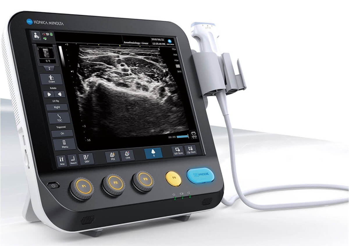
| Content | Konica Sonimage MX1 Ultrasound
Konica Sonimage MX1 Ultrasound Point-of-Care system inherits a DNA of innovation from the higher-end model SONIMAGE HS1. The system delivers advanced technologies that ensure superior image quality and efficient workflow – all with exceptional value.Intuitive Operation
Konica Sonimage MX1 Ultrasound Customizable touchscreen with the five most frequently used keys allows you to facilitate superior workflow and improve efficiency and throughput.Cradle (Cable stress free)
Cradle, a mounting platform for MX1, allows you to hand-carry the system without insertion and removal of AC power/USB cable. It can be mounted on the cart with VESA and enables you to charge the battery when you put the system back.MPA, a function unique to Konica Minolta, enables you to change multiple image parameters like frequency and turn trapezoid on in conjunction with depth change. It reduces unnecessary key strokes to optimize the image and increase throughput.Dual Sonic, Konica Minolta’s unique technology, uses a unique transmitting algorithm which can transmit two waveforms depending on the focusing depth.In combination with T2HI technology, it forms the high quality of THI signal around the center of ultrasound beam in the receiving area. As a result, it suppresses the acoustic noise to ensure the optimum image from deep area to superficial.Anytime, Anywhere for anyoneThe SONIMAGE MX1 Point-of-Care system inherits a DNA of innovation from the higher-end model SONIMAGE HS1. The system delivers advanced technologies that ensure superior image quality and efficient workflow - all with exceptional value. Workflow EfficiencyIntuitive Operation
Customizable touchscreen with the five most frequently used keys allows you to facilitate superior workflow and improve efficiency and throughput. Easy and QuickCradle (Cable stress free)
Cradle, a mounting platform for MX1, allows you to hand-carry the system without insertion and removal of AC power/USB cable. It can be mounted on the cart with VESA and enables you to charge the battery when you put the system back. | Acuson Cypress PLUS UltrasoundAcuson Cypress cardiovascular system PLUS delivers advanced ergonomics and superior performance in a highly portable package. TheCypress System PLUSis a highly miniaturized, all digital, phased array echocardiography system that provides complete studies and outstanding images – even on patients who are difficult to image.The Cypress PLUS transitions easily from your small private office or hospital echo lab to the operating theatre or your patient’s bedside. It’s ultrasound where you need it.The Cypress PLUS delivers high performance in a complete range of cardiovascular capabilities.Designed with advanced ergonomics in mind, the Cypress system PLUS streamlines your workflow and increases your productivity.If you already own a Cypress system, keeping technologically current is simple with the Cypress Upgrade program.The Cypress system is the smallest, lightest, high performance, phased array echo system ever built. Weighing in at just 19 pounds, with digital storage capacity for over 100 studies and network capabilities to send studies from remote locations, clinicians can perform complete echocardiography studies from almost anywhere. TheCypress system was designed for portability, durability and reliability, and to withstand the rigors of constant transportation. Internal system interconnects are minimized. Key components are shock mounted and the entire system sits inside a robust chassis. From hospital to office or from airplane to mountain village, the Cypress system will perform.Full Range of Capabilities
The Cypress system is a complete, full-featured echo cardiography system that provides complete studies and outstanding image quality even on the most difficult to image patients. It offers a full range of features including:Acuson Cypress PLUS Ultrasound– 2D with Harmonics
– Patient Report with audio capture
– M-Mode
– Embedded DICOM conformance
– Color PW and CW Doppler
– High frame rate color flow imaging
– Stress echo
– Network output
– Vascular imaging
– Comprehensive calculations package
– Contrast agent imaging
– USB peripheral support (PC printer connectivity)Physical Characteristics
• Single unit houses ultrasound system with control panel, flat panel display and MO drive
• Weight: 8.6 Kg (19 pounds)
• Height: 35.6 cm (14 in)
• Width: 40.6 cm (16 in)
• Depth: 20.3 cm (8 in) – with keyboard folded up 34.3 cm (13.5 in) – with keyboard down
• Heat dissipation: 490 BTU/hour (nominal)User Interface
• Fold-up control panel for portability
• Direct access to system functions through dedicated keyboard controls and multifunctional soft keys
• Easy-access main controls, including multifunctional trackball, enter and select keys, for minimum hand movement while controlling the system
• Adjustable trackball speed
• Functional grouping of keys, rotational knobs, switches and sliding TGC controls for direct access to critical functions
• Tactile feedback on control activation
• Backlighting of control panel labels
• Mode-dependent soft keys
• User-interface tools
• Pre-defined and user-entered labeling textMonitor
• 10.4 inch/26.4 cm flat panel active matrix display screen
• Protective anti-glare, anti-reflective lensHard Drive
• Internal hard drive for image and data storage
• Internal storage of approximately 50 patient studies (25 – 30 loops per study)Transducer Ports
• One transducer connector (type IEC601BF)Operating/Display Modes
• 2-D, fundamental and harmonic imaging
• Color Doppler Velocity (CDV)
• Color Doppler Energy (CDE)
• M-mode, vertical split screen display with 2-D
• Pulsed Wave (PW) Spectral Doppler
• Continuous Wave (CW) Spectral Doppler
• Duplex Doppler (combined 2-D and Spectral Doppler display)2D Calipers – Generic Measurements and Calculations
• Measurements and calculations in 2D, M-mode, color Doppler, PW and CW spectral Doppler modes
• Basic measurements and report calculations
• Comprehensive patient calculations report
• On-screen “mini-reports”
• Complete or configured patient report
• Cardiac package
• Carotid package
• Arterial package
• Venous package
• Abdominal package
• User configurable Sites
• Selectable menu position
• Four cursor types
• Angle correction
• Distance
• Area
• Slope
• Time
• Velocity
• Velocity/pressure
• Mean velocity/pressure gradient
• Velocity Time Integral
• Pressure halftime
• Continuity Equation
• Ejection Fraction
• Left Ventricular Volumes, including End-Systolic and End-Diastolic volumes
• Cardiac output
• Cardiac index
• Fractional area changeCine Review
• 2D Cine memory up to 15 seconds of history
• Color Doppler cine memory up to 15 seconds of history
• M-mode strip cine along with PW and CW Doppler mode
• strip cine memory determined by sweep speed (selectable by the user) 10, 20, 30 or 40 secondsTransducer Technology
• 3V2c: 2-4 MHz wideband phased array
• 7V3c: 3-7 MHz wideband phased array
• 7L3: 5-7 MHz wideband linear array (Vascular)
• 4C1: 2-4 MHz wideband curved linear array (Abdominal)
• V5Ms: 3-6 MHz wideband phased array (micro-pinless (MP) connector) | CLASSYS ULTRAFORMER III UTR3
You’ve heard about it, now see and feel the difference for yourself.Ultraformer III is the leading technology for facial lifting, tightening and contouring. Now with 7 interchangeable cartridges that offer your patients another dimension in non-surgical face and body treatments.Ultraformer III is widely used with over 10 million treatments performed worldwide. It has become the treatment of choice for patients who are not yet indicated or not willing to undergo surgery. This fills a huge gap in the market, and captures an audience of people from 30 years of age all the way to 80.ULTRAFORMER III FEATURESLess discomfort associated with treatments
Patented transducer design for more efficient energy delivery
Dual hand pieces with automatic recognition for ease of use
Faster shot and treatment speed to save time
Easy, user-friendly design and interface
Shot count information and voice assistance
Backed by clinical papersThe CLASSYS ULTRAFORMER III UTR3 advantage with slim transducer designimproves operator visibility
supports very narrow focus and precise treatment
fits facial contoursFast elliptic transducersmore efficient treatment timesUltraformer III’s 7 clip-in transducer cartridges project uniform ultrasound beams directly to multiple layers underneath the skin to promote tighter collagen formation, tissue contraction and reduction in the volume of adipocytes that form bulging areas of tissue.Patients notice an immediate benefit in improved skin tone and texture, with ongoing improvements over 3 to 6 months
Traditionally difficult areas such as eye lifting are made easier with Ultraformer III’s specialised cartridges. It is possible for patients to experience up to 6mm of eyebrow lift, depending on their particular situation.Ultraformer III’s 7 clip-in transducer cartridges project uniform ultrasound beams directly to multiple layers underneath the skin to promote tighter collagen formation, tissue contraction and reduction in the volume of adipocytes that form bulging areas of tissue.Patients notice an immediate benefit in improved skin tone and texture, with ongoing improvements over 3 to 6 months
Traditionally difficult areas such as eye lifting are made easier with Ultraformer III’s specialised cartridges. It is possible for patients to experience up to 6mm of eyebrow lift, depending on their particular situation. | GE Voluson i Portable 3D/4D UltrasoundFeatures* Lightweight and portable
* Battery operation
* Real-Time 4D
* 3D Multiplanar Display
* 3D Power Doppler
* Automatic Optimization (AO)
* Speckle Reduction Imaging (SRI) and CrossXBeam deliver advanced computational power, allowing for simultaneous processing of CrossXBeam CRI and SRI together – enabling added speckle reduction, contrast resolution and image clarity.
* HD-Flow uses a bi-directional Doppler feature to achieve a more sensitive vascular study and reduce overwriting.
* Volume Contrast Imaging (VCI) allows for image quality in either a single plane or all three planes of a volume acquisition
* Spatio-Temporal Image Correlation (STIC) captures a full virtual fetal heart cycle in real time, and the volume can be saved for offline analysis.
* Tomographic Ultrasound Imaging (TUI) makes analysis and documentation of dynamic studies easier with a simultaneous view of multiple parallel slices of a volume data set.
* Virtual Organ Computer-aided AnaLysis (VOCAL) is a semi-automated volume measurement tool that utilizes computer technology to provide accurate volume calculations.Comes with:GE Voluson i Portable 3D/4D Ultrasound* 1x Voluson i Portable 3D/4D Ultrasound
* 1x RAB2-5RS (2-5MHz) 3/4D Convex Abdominal Probe
* 1x E8C-RS (4-10MHz) Wideband Microconvex Endocavity (Vaginal) Probe
* 1x RIC5-9-RS (5-9MHz) 2D/3D RealTime 4D Endocavitary ProbeThe GE Voluson i ultrasound machine is primarily used for OB-GYN applications, but is versatile and powerful enough to be used for a variety of other applications as well. The Voluson i ultrasound machine carries the power of the GE Voluson line with access to premium 2D and 4D imaging in a portable form factor. The GE Voluson i provides the extraordinary image quality needed to confidently scan for women’s health. The GE Voluson i portable ultrasound machine offers features such as speckle reduction imaging and CrossXBeamCRI, which both lend to the incredible image clarity and quality of this system. The Voluson I portable ultrasound system is known for ease of use, allowing the operator to scan patients faster and more efficiently with better accuracy. | DOUBLO GOLD HIFU SYSTEMWhy choose Doublo HIFU System?
HIFU (High-Intensity Focused Ultrasound System) is a highly precise medical procedure using high-intensity focused ultrasound to heat and destroy the pathogenic tissue rapidly. The earliest widespread use of HIFU was as a treatment for prostate cancer.HIFU hybrid system for Face & Body
Doublo Gold is the ultimate HIFU system for precise Face Lifting & Body Contouring. Offering the fastest treatment by Two-way round shooting and Auto Shot mode.Non Invasive – No Pain – No Bleeding – No Downtime
Faster shot speed : Efficacy improved by repetitious shooting with 300 shots in 8 minutes. Entertain more patients with a faster treatment procedure.Multiple treatment areas
By treating the upper dermis with wound healing effect, facial pores and skin tone is improved as the fibroblast is rebuilt and collagen is boosted.Dermal tissue elasticity and collagen increase as fibroblast is rebuilt by wound healing effect from damaging sub-cutaneous tissue and derma layer with thermal coagulation.
Skin tightening to reduce wrinkle and increase lifting effect is done by creating thermal coagulation at subcutaneous fat and SMASDOUBLO GOLD HIFU SYSTEM Highly Precise, Non-Surgical, Medical Procedure System for Professionals. Doublo HIFU’s Speed is Innovative Performance! Treatment time is dramatically reduced with Hironic’s High-capacity Technology and with Doublo HIFU System you can treat twice the amount of patients than beforeDOUBLO GOLD HIFU SYSTEM Accessories: Power cord for cart
Clear plastic tray insert
Large washer and small lock/spring washer
2x black attachements
2x treatment parameters guide
Simple user manual
Full user manual + CDHighly Precise, Non-Surgical, Medical Procedure System for Professionals. Doublo HIFU’s Speed is Innovative Performance! Treatment time is dramatically reduced with Hironic’s High-capacity Technology and with Doublo HIFU System you can treat twice the amount of patients than beforeNew Standard of
MFU System
1.No. 1 MFU proven by the patents2.MFU proven by cumulative sales and clinical cases3.Fastest treatment by Two-way round shooting and Auto Shot mode | Samsung Medison SonoAce R3 Ultrasound Machine
SonoAce R3 is a portable color ultrasound system with comprehensive diagnostic capabilities. Full Spectrum Imaging™ (FSI™) combines with Speckle Reduction Filter™ (SRF™) to deliver superior quality images, even in the challenging circumstances. The compact and user-friendly design features of the SonoAce R3 ensure portability and manoeuvrability in a lightweight unit. Sporting a 15-inch LCD monitor with Full-Featured Workflow, the SonoAceR3 offers your organisation flexibility, without compromising on precision and efficiency.Slim and Compact DesignThe compact design of SonoAce R3 offers Various-Featured Workflow with a thumbnail menu and shortcut, together with functional key groupings for increased efficiency. The user-friendly design combines a wide-view 15-inch LCD monitor with backlit control keys, customisable measurement package and body markers. The SonoAce R3 features 2 probe ports, 3 USB ports, rapid boot up capability and DICOM 3 compatible image filing. This slim, lightweight unit also has a convenient carrying handle for portability and flexibility.Samsung Medison SonoAce R3 Ultrasound MachineFull Spectrum Imaging™ (FSI™) produces the penetration capabilities associated with the lower frequencies, while still maintaining the fine pixel uniformity associated with the higher frequencies. This combination consistently delivers superior quality images, even in the challenging diagnostic circumstances. The resulting clarity delivers improvements in accuracy and efficiency for greater confidence in your diagnostic procedures. Harmonic Imaging is a technique that records higher frequency information, resulting in greater resolution and fewer artifacts.Synthetic Aperture
The physical radio channel may present some limits to imaging which can be overcome by using Synthetic Aperture Control software. This method uses information from diverse sources to form a more better picture of the area under examination. It creates one single scan line by performing the TX and RX twice to create enhanced penetration and resolution. Synthetic Aperture Control technology delivers a comprehensive picture and to aid accurate diagnosis.QuickScan
QuickScan has been designed to increase workflow efficiency by automatically optimising key imaging parameters at the touch of a button. The resulting increase in efficiency allows your organisation to offer patients an improved service with faster and more accurate diagnoses. This reliable and safe system helps your clinic or hospital to deliver a superior patient experience.Features
Full Spectrum ImagingTM
Quick ScanTM
Color and Power Doppler
Harmonic imaging
Freehand 3D
3D Multi Planar Imaging
Sonoview Image Management
PW Doppler
15″ LCD monitor
Two probe ports
Lightweight |




Reviews
There are no reviews yet.