| Description | - Superior Image Quality (T2HI)
- Greater portability
- Innovative design
- Intuitive user interface
- Simple Needle Visualization (SNV) technology
| Function • Gravity Sensor: You can operate the ultrasound system either in transversal layout or vertical layout.• Grid for estimation: With grid on the image area, you can easily estimate the size of the target substance like follicle fertilized egg etc. | The GE Vivid q portable ultrasound machine is a more powerful version of the Vivid i that adds further quantitative features including ICE, Auto EF, AFI, TVI,TT, TSI, Blood Flow and Blood Flow imaging. The Vivid q is the pinnacle of GE’s portable cardiovascular ultrasounds. | The SonoSite Titan is the earliest version of the PoC dedicated portable ultrasound machine from Sonosite. Though its form factor is compact, it provides high resolution imaging, rapid responsive time, and supports a broad range of applications including cardiac. | GE Voluson 730 Expert is among the top-regarded ultrasounds in the industry. With up to 40 volumes per second rate of acquisition and construction of volumetric images in real time. The 730 Expert is loaded with the newest ultrasound technology including: CrossXBeam, Speckle Reduction Imaging, Tomographic Ultrasound Imaging (TUI), Spatio-Temporal Image Correlation (STIC), and much more. | The more patients you see, the more you need Voluson E8.
The Voluson E8 ultrasound system is designed to keep pace with busy practices that conduct a wide range of Women’s Health exams, from routine scanning to complex assessments.
The imaging excellence of Radiance System Architecture – your diagnostic confidence will be enhanced by the extraordinary image clarity and speed of Radiant System Architecture. |
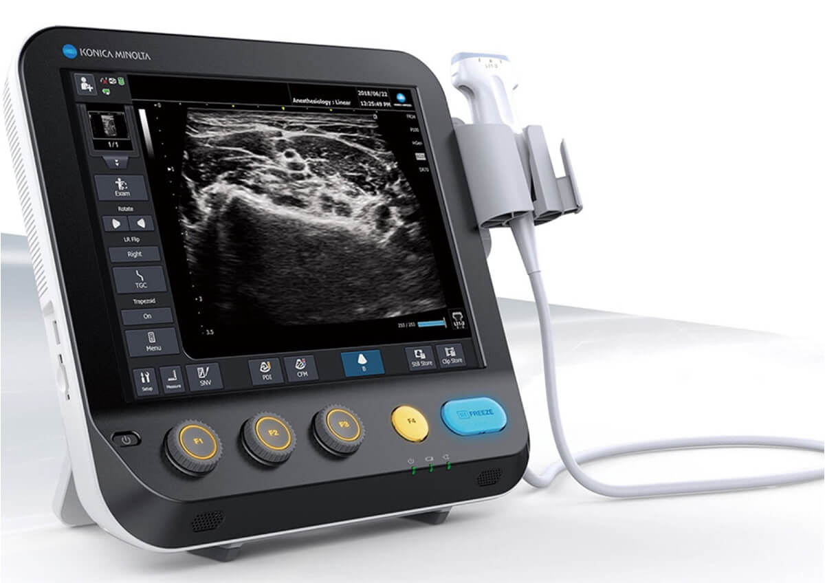
| Content | Konica Sonimage MX1 Ultrasound
Konica Sonimage MX1 Ultrasound Point-of-Care system inherits a DNA of innovation from the higher-end model SONIMAGE HS1. The system delivers advanced technologies that ensure superior image quality and efficient workflow – all with exceptional value.Intuitive Operation
Konica Sonimage MX1 Ultrasound Customizable touchscreen with the five most frequently used keys allows you to facilitate superior workflow and improve efficiency and throughput.Cradle (Cable stress free)
Cradle, a mounting platform for MX1, allows you to hand-carry the system without insertion and removal of AC power/USB cable. It can be mounted on the cart with VESA and enables you to charge the battery when you put the system back.MPA, a function unique to Konica Minolta, enables you to change multiple image parameters like frequency and turn trapezoid on in conjunction with depth change. It reduces unnecessary key strokes to optimize the image and increase throughput.Dual Sonic, Konica Minolta’s unique technology, uses a unique transmitting algorithm which can transmit two waveforms depending on the focusing depth.In combination with T2HI technology, it forms the high quality of THI signal around the center of ultrasound beam in the receiving area. As a result, it suppresses the acoustic noise to ensure the optimum image from deep area to superficial.Anytime, Anywhere for anyoneThe SONIMAGE MX1 Point-of-Care system inherits a DNA of innovation from the higher-end model SONIMAGE HS1. The system delivers advanced technologies that ensure superior image quality and efficient workflow - all with exceptional value. Workflow EfficiencyIntuitive Operation
Customizable touchscreen with the five most frequently used keys allows you to facilitate superior workflow and improve efficiency and throughput. Easy and QuickCradle (Cable stress free)
Cradle, a mounting platform for MX1, allows you to hand-carry the system without insertion and removal of AC power/USB cable. It can be mounted on the cart with VESA and enables you to charge the battery when you put the system back. | SIUI CTS-800 Veterinary Ultrasound Machine with Linear Rectal Probe
This small and convenient “Palm-Sized” portable veterinary ultrasound scanner fits in the palm of your hand, and is mostly used for farm animals. Its long battery life and waterproof features makes it the perfect ultrasound scanner for veterinarians on the field.The screen of the SIUI CTS-800V will rotate horizontally or vertically, according to how you are holding it.Gravity Sensor: You can operate the ultrasound system either in transversal layout or vertical layout. It can automatically adapt to two different layouts when you rotate it.Grid for estimation: With grid on the image area you can easily estimate the size of the target substance like follicle fertilized egg etc.SPECIFICATIONS SIUI CTS-800 Veterinary Ultrasound Machine with Linear Rectal ProbeMonitor: 7-inch WVGA LCD monitorWeight: 0.8kg Environmental rating: IP54 (main unit) IP67 (probe head) Battery: 3 hours operation time Display mode: B 2B ZOOM B B/M M Depth: Max to 250mm B gain: 0-100 Image storage: 256 frames Cine loop: 256 frames Probe: L50/7.5 MHz linear rectal probe (standard) L65/5.0 MHz linear rectal probe (optional) R20/5.0 MHz micro convex probe (optional)
– Main unit with 7-inch LCD monitor.
– 1 Probe connector.
– 1 Li-ion battery. 3 hours operation time.
– 1 Adaptor.
– 1 Car Charger.
– Belt and sun shield.
– 1 USB port (Mini).
– Carrying case.PROBES– Rectal Linear L7FVC (6.5/7.1/7.5/8.3/9.0MHz).
– Rectal Linear L5FVC (4.0/4.7/5.0/5.5/6.2MHz).
– Backfat Linear L1FC (150mm, 2.5/3.0/3.5/4.0/5.0MHz).
– Convex C3FC (2.5/3.0/3.5/4.0/5.0MHz).
– Microconvex C5FC (3.5/4.0/5.0/6.5/7.1MHz).CTS-800 is a palm-sized veterinary ultrasound system. Features Weight: 0.8kg Environmental rating: IP54 (main unit) IP67 (probe head) Battery: 3 hours operation time Display mode: B 2B ZOOM B B/M M Depth: Max to 250mm B gain: 0-100 Image storage: 256 frames Cine loop: 256 frames Probe: L50/7.5 MHz linear rectal probe (standard) L65/5.0 MHz linear rectal probe (optional) R20/5.0 MHz micro convex probe (optional) | GE Vivid Q Ultrasound Machine
The powerful GE Vivid Q ultrasound machine is the ultimate lightweight, compact high-performance cardiovascular system for clinicians needing a machine that combines user efficiency and reliable performance. This unique system performs consistently thanks to its TruScan architecture and can support up to 17 probes. The GE Vivid Q is recognized for its premium image quality, advanced image management, and high-performance quantification tools.This advanced GE ultrasound delivers improved patient care and enhanced user productivity with the new M4s-Matrix array technology transducer, which provides greatly enhanced image quality in electronic beamforming, raw data image acquisition and archiving, optimal onboard and post processing technology.This ultrasound system provides high clinical values for quantification and review of stored images, making it one of today’s most sought-after models. Some of its powerful features include compatibility with EchoPac clinical workstation and DICOM networking, speckle tracking strain, integrated stress echo, quantification tools, and reporting capabilities. Also of note is one of the Vivid Q’s best new features, which is its TEE transducer capability.A one-touch button optimization provides premium image quality for workflow efficiency, and maintains AFI- automatic function imaging with complete wallFeatures- Advanced QScan Imaging (incl TSI)
- AdvStress
- ATO
- Auto EF
- Automated Function Imaging
- Blood Flow Imaging
- CardiacApp
- Cardio Lab
- Color Doppler
- CW Doppler
- DICOM Networking
- DICOM: Print
- DICOM: Worklist
- DVD/RW
- ECG
- ECG Cable
- EchoDicom
- EchoPAC
- Evue
- ICE Imaging
- Intima Media Thickness (IMT)
- LVO Contrast
- M Mode
- M4S
- MPEGvue
- PW Doppler
- Small Depth
- TEE
- THI
- Tissue Harmonics Imaging (THI/NTHI)
- Tissue Velocity Imagine & Tissue Tracking
- TSI
- VascAbdContrast
- VascularApp
- VirtualPrinter
- Wide Aperture
GE Vivid Q FeaturesThe GE Vivid Qis a Portable Laptop-style Ultrasound that can be used to help diagnose cardiovascular anatomy. Features and tools based on the previous Vivid 7 and the Vivid E9 have been added to the new lightweight Vivid Q, mainly the M4S-RS transducer that allows for 3D image focusing and enables a higher level of image quality. The Vivid Q ultrasound is versatile and can additionally be used for Cardiac, vascular, Abdominal, Pediatric, Obstetrics, and Emergency medicine. The Vivid Q Ultrasound comes with a 15-inch color LCD monitor that can display a variety of imaging modes including; ICE, Auto EF, AFI, TVI, TT, TSI, and Blood Flow. Additional the Ultrasound system is comparable with 17 different transducers and probes to fit every imaging need. - Auto EF - Automated ejection fraction measurement program designed specifically for the GE Vivid q.
- Tissue Synchronization Imaging (TSI).
- Intracardiac Echo (ICE).
- 16 different compatible probes.
- Coded Octave imaging - enhances image color flow and Doppler imaging.
| Sonosite Titan Ultrasound Machine
The SonoSite Titan Portable Ultrasound is SonoSite’s second generation portable ultrasound system. This system improved upon the technology of the SonoSite 180 with a larger screen and keyboard packed into a laptop style system. This system is well suited for Vascular access, Musculoskeletal, Anesthesiology, Internal Medecine, Emergency rooms and Family Physicians. Veterinary configurations are available. Veterinary configurations are available.8.4″ LCD monitor, 2 probe ports, Vascular, OB/GYN, Cardiac reports, Soft keys, Split screen, Zoom, Storage via 64 or 256 MB flash cards, 220 image CINE loop, USB & Ethernet image transfer, Volume calculations via diameters, THI [Tissue Harmonic Imaging], SonoCalc [IMT analysis, PC-based], DICOM, S-video (in/out), RGB, Composite video.
Dimensions: 3″ (H) x 10.9″ (W) x 11.9″ (D) Weight: 7.7 lbs Manufacturer
Sonosite Application
Cardiology, Endocrinology, Family Practice, Gastroenterology, Internal Medicine, Nephrology, Neurology, OB-GYN, Orthopedics, Otorhinolaryngology, Pain Management, Surgery, Urology, Vascular Probes - C8 – prostate – 8-5 MHz 8-mm broadband intracavitary array – 8.5 cm – Available – MicroMaxx/ TITAN
- C11 – abdominal/ pediatric cardiology/ neonatal/ vascular – 8-5 MHz 11-mm broadband curved array – 10 cm – N/A – TITAN
- C15 – adult cardiology/ abdominal – 4-2 MHz 15-mm broadband tightly curved array – 25 cm – N/A – TITAN
- C60 – abdominal/ obstetrics/ gynecology – 5-2 MHz 60-mm broadband curved array – 22 cm – Available – TITAN
- ICTe – obstetrics/ gynecology – 8-5 MHz 11-mm broadband tightly curved array – 10 cm – Available – MicroMaxx/ TITAN
- L25 – nerve/ musculoskeletal/ vascular and superficial – 10-5 MHz 25-mm broadband linear array – 6 cm – Transverse Guide Available – TITAN
- L38 – small parts/ breast/ vascular/ nerve/ musculoskeletal/ superficial – 10-5 MHz 38-mm broadband linear array – 6.5 cm – Available – TITAN
Options - Extended cardiac package
- Pulsed wave (PW) doppler
- Continuous wave (CW) doppler
- Tissue Harmonic Imaging
Accessories - Cart
- Black and White Video Printer
- DVD Recorder
The SonoSite Titan is the earliest version of the PoC dedicated portable ultrasound machine from Sonosite. Though its form factor is compact, it provides high resolution imaging, rapid responsive time, and supports a broad range of applications including cardiac. | GE Voluson 730 Expert Ultrasound MachineGE Voluson 730 Expert, 3D/4D Realtime Ultrasound System Color Doppler, PW Doppler, Tissue Harmonics, STIC, VOCAL DICOM, M-mode, Angio, ATO, TUI, SonoView II, Crossbeam (CRI) MO Drive, Cineloop, High Resoluotion Interlaced Monitor Probe Options: RAB4-8 4D Convex RIC5-9 4D Transvaginal AB2-7 2D Convex IC5-9 2D Transvaginal SP6-12 2D Linear PA2-5 Cardiac Sector
Two Probes included:- RAB4-8L – Live 4D Abdominal Transducer
- RIC5-9 – Live 4D Transvaginal Transducer
Options:
About the Voluson 730:GE Voluson 730 Expert Ultrasound MachineWith GE Voluson 730, you can now acquire and construct volumetric images in real time – up to 40 volumes per second. The system allows you to explore images in any plane to reveal the smallest details with stunning clarity and apply sophisticated analytical tools to answer virtually any of your clinical questions. Volume Ultrasound not only improves your 3D/4D capabilities, it enhances your 2D imaging as well, for even greater diagnostic confidence in your obstetrical, gynecologic, breast and general imaging studies.The used GE Voluson 730 Expert was among the top-rated and biggest breakthrough ultrasounds in the industry. With up to 40 volumes per second rate of acquisition and construction of volumetric images in real time. The 730 Expert is loaded with the newest ultrasound technology including: CrossXBeam, Speckle Reduction Imaging, Tomographic Ultrasound Imaging (TUI), Spatio-Temporal Image Correlation (STIC), and much more.The Voluson 730 Expert is the more advanced version of the Voluson 730 Pro and was replaced by the GE Voluson e8. Compared to the 730 Pro, the Voluson 730 Expert features a touchscreen interface, higher frame rates in 2D and 4D, and more advanced imaging options such as STIC and TUI.Providian Medical Voluson 730 Expert Review:The Voluson 730 is a workhorse of a 4D ultrasound machine. It remains a popular system in many OB/GYN/Women’s Health offices, as well as 4D “Babyface” studios around the world. It is a durable machine with very good image quality for all women’s health needs. | GE Voluson E8 Ultrasound Machine15’’ LCD monitor [Rev BT06], 10.4” touchscreen, 3 Active Probe Ports, Floating Keyboard: Interactive back-lighting, Beta-View CrossXBeam, Virtual Convex, Extended View, ATO [Automatic Tissue Optimization], Coded Harmonic Imaging, High Resolution Zoom, Inversion Real-time automatic Doppler, OB/GYN; Vasc. Cardiac; Abd; Small-Parts; Urology; Ped; Ortho; Neuro reporting, Multigestational Calculations, HPRF [High Pulse Repetition Frequency], SonoVCAD [Matrix array volume technology], TruScan [On-board archive], VCI [Volume Contrast Imaging], SonoAVCfollicle [Sonography-based Automated Volume Count follicle], SonoVCADheart [Sonography-based Volume Computer Aided Display], TUI [Tomographic Ultrasound Imaging], CE [Coded Excitation], XTD, SRI III [Speckle reduction imaging], CRI [Compound Resolution Imaging], DICOM, Ethernet port, Video Out, 6 USB ports, Integrated HDD (160 GB) and DVD/CD-RW.
3D/4D, Endocrinology, Family Practice, Gastroenterology, Internal Medicine, Nephrology, Neurology, OB-GYN, Orthopedics, Otorhinolaryngology, Pain Management, Urology, Vascular Probes GE Voluson E8 Ultrasound Machine - 11L-D Linear Array
- 3S-D Cardiac Sector
- 4C-D Convex Array
- 9L-D Linear Array
- AB2-7 Convex
- IC5-9-D Intercavity
- M6C-D Convex
- P2D CWD Non-Imaging
- P6D CWD Non-Imaging
- PA6-8-D Cardiac
- RAB2-5-D 3D/ 4D Convex
- RAB4-8-D 3D/4D Convex
- RIC5-9-D 3D/4D Intracavity
- RIC6-12-D 3D/4D Intracavity
- RNA5-9-D 3D/4D Convex
- RRE6-10-D Endocavitary Micro-Convex
- RSP6-16-D 3D/D Linear
- SP10-16-D Linear
- 19″ monitor
- 4D Real Time
- CFM Doppler Mode
- Coded Contrast Imaging
- CrossBeam
- DICOM 4D
- HD flow
- Power Doppler Mode
- SonoVCAD
- STIC (Spacial-Temporal Image Correlation)
- T.U.I (Tomographic Ulrasound Imaging)
- VOCAL II VCI (Volume Contrast Imaging)
- Black and white printer
- Color printer
- DVD-Recorder
As your patient volume increases and your clinical cases become more complex, you will require an ultrasound system that enables you to deliver truly exceptional care – confidently and efficiently – every time. The Voluson E8 ultrasound system is designed to keep pace with busy practices – handling routine to complex women’s health exams with ease and precision. The premium image quality of the Voluson E8 combined with access to advanced capabilities, delivers the level of performance required by you and your patients. |




Reviews
There are no reviews yet.