| Description | All necessary adjustments and operations are realized automatically.
By touching the mode and measurement button, semi-automatic measurement also achieved.
This function supports easy workflow for blood vessels and blood flow volume measurements. | The more patients you see, the more you need Voluson E8.
The Voluson E8 ultrasound system is designed to keep pace with busy practices that conduct a wide range of Women’s Health exams, from routine scanning to complex assessments.
The imaging excellence of Radiance System Architecture – your diagnostic confidence will be enhanced by the extraordinary image clarity and speed of Radiant System Architecture. | Acuson sequoia 512 ultrasound click images to enlarge item description acuson sequoia 512 ultrasoundsold as picturedincludes: 2 probes, 4c1 convex & 6l4 linearsony up 895 md printerunit will be cleaned, function checked and certified for electrical safety. Checked, cleaned, certified for electrical safetyguaranteed working when arrives on the guarantee working we need to be notified in 48 hrs. And the seals must be intact. | The GE Vivid q portable ultrasound machine is a more powerful version of the Vivid i that adds further quantitative features including ICE, Auto EF, AFI, TVI,TT, TSI, Blood Flow and Blood Flow imaging. The Vivid q is the pinnacle of GE’s portable cardiovascular ultrasounds. | The Sequoia system’s advanced technologies are designed to make image acquisition easier, reduce exam time and increase overall efficiency. Along with many other included features, the Sequoia system offers Native, a patient specific imaging which adapts both phase and amplitude information of each unique patient’s properties which delivers a consistently better image across a broad range of patients. | Product Features:- Exclusive Raw Data Digital acquisition and storage
- Color Doppler, Spectral Dopler, B, M, Triplex, 3-D
- 150+ Frames per Second TrueDigital imaging
- All digital video clip and still image capture (4,000)
- On-board patient, image and reporting archive
- CD-ROM disk burner included, plus PCMCIA and USB
- Raw Data manipulation on image recall for Post Exam:
- Anatomic M-mode creation and adjustment
- Gain, magnification, and colorization control
- Playback speed, sweep speed, angle, baseline, and more…
- 3-D Digital processing and manipulation o Clip and still measure-annotate-enhance-store new
- DICOM 3.0 capable, plus VetPACS all Digital interfacing
- Angio
- ATO
- TMI
- AMM
- Virtual Convex
- PW/CW Doppler
- ECG/AUX
|
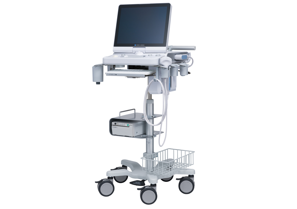
| Content | Konica Sonimage HS2 Ultrasound
Konica Sonimage HS2 Ultrasound is a Point-of-Care portable ultrasound system designed to support a wide range of applications and patient types. Konica Minolta’s advanced technology features provide high resolution image quality and efficient workflow for daily clinical practice.Konica Sonimage HS2 Ultrasound Unique Nanofabrication Technology, combines both material and machining expertise, to produce broad frequency linear probe “L18-4”. It offers both high sensitivity as well as greater penetration.T2HI (Traid Tissue Harmonic Imaging)
Dual Sonic Technology
By combining T2HI and Dual Sonic, high quality THI signal is formed around the center of ultrasound beam in receiving area.
As a result, it suppresses the acoustic noise and ensure the optimum image from deep to superficial structure.iXRET
The SONIMAGE HS2 achieves higher resolution and faster frame rates by utilizing Konica Minolta’s unique technologySNV (Simple Needle Visualization)
The SONIMAGE HS2 automatically detects needle insertion.
This funciton can be used both ‘In plane’ and ‘Out of planeEfficient Workflow for Daily Clinical Practice The SONIMAGE HS2 delivers intuitive workflow by customizing 8 physical buttons and touch panel. MPA (Multi Parameter Adjuster) MPA enables to change multiple image parameters like frequency change and turning trapezoid on in conjunction with depth change. Vascular NAVI
All necessary adjustments and operations are realized automatically.
By touching the mode and measurement button, semi-automatic measurement also achieved.
This function supports easy workflow for blood vessels and blood flow volume measurements. The SONIMAGE HS2 offers the unique capability to write text and draw lines and figures using fingers on the screen. This is an excellent tool for training and communication with patients. | GE Voluson E8 Ultrasound Machine15’’ LCD monitor [Rev BT06], 10.4” touchscreen, 3 Active Probe Ports, Floating Keyboard: Interactive back-lighting, Beta-View CrossXBeam, Virtual Convex, Extended View, ATO [Automatic Tissue Optimization], Coded Harmonic Imaging, High Resolution Zoom, Inversion Real-time automatic Doppler, OB/GYN; Vasc. Cardiac; Abd; Small-Parts; Urology; Ped; Ortho; Neuro reporting, Multigestational Calculations, HPRF [High Pulse Repetition Frequency], SonoVCAD [Matrix array volume technology], TruScan [On-board archive], VCI [Volume Contrast Imaging], SonoAVCfollicle [Sonography-based Automated Volume Count follicle], SonoVCADheart [Sonography-based Volume Computer Aided Display], TUI [Tomographic Ultrasound Imaging], CE [Coded Excitation], XTD, SRI III [Speckle reduction imaging], CRI [Compound Resolution Imaging], DICOM, Ethernet port, Video Out, 6 USB ports, Integrated HDD (160 GB) and DVD/CD-RW.
3D/4D, Endocrinology, Family Practice, Gastroenterology, Internal Medicine, Nephrology, Neurology, OB-GYN, Orthopedics, Otorhinolaryngology, Pain Management, Urology, Vascular Probes GE Voluson E8 Ultrasound Machine - 11L-D Linear Array
- 3S-D Cardiac Sector
- 4C-D Convex Array
- 9L-D Linear Array
- AB2-7 Convex
- IC5-9-D Intercavity
- M6C-D Convex
- P2D CWD Non-Imaging
- P6D CWD Non-Imaging
- PA6-8-D Cardiac
- RAB2-5-D 3D/ 4D Convex
- RAB4-8-D 3D/4D Convex
- RIC5-9-D 3D/4D Intracavity
- RIC6-12-D 3D/4D Intracavity
- RNA5-9-D 3D/4D Convex
- RRE6-10-D Endocavitary Micro-Convex
- RSP6-16-D 3D/D Linear
- SP10-16-D Linear
- 19″ monitor
- 4D Real Time
- CFM Doppler Mode
- Coded Contrast Imaging
- CrossBeam
- DICOM 4D
- HD flow
- Power Doppler Mode
- SonoVCAD
- STIC (Spacial-Temporal Image Correlation)
- T.U.I (Tomographic Ulrasound Imaging)
- VOCAL II VCI (Volume Contrast Imaging)
- Black and white printer
- Color printer
- DVD-Recorder
As your patient volume increases and your clinical cases become more complex, you will require an ultrasound system that enables you to deliver truly exceptional care – confidently and efficiently – every time. The Voluson E8 ultrasound system is designed to keep pace with busy practices – handling routine to complex women’s health exams with ease and precision. The premium image quality of the Voluson E8 combined with access to advanced capabilities, delivers the level of performance required by you and your patients. | Siemens / Acuson Sequoia 512 Ultrasound Machine
The ACUSON Sequoia 512 ultrasound is an ultra-premium system that utilizes revolutionary technology, dedicated to the advancement of diagnostic imaging. It represents some of the most advanced technology in ultrasound imaging. The Acuson Sequoia 512 has unparalleled imaging performance, greater image content, enhanced detail, superior contrast resolution & penetration, and excellent color Doppler sensitivity.One of the best selling ultrasounds in the United States, the Sequois 512 provides superior imaging with Native Patient Specific Imaging that detects, measures and adapts (in real time) to each patient’s individual acoustics.Performance:Reduce image optimization time by up to 80% and allow for an easier, faster and more confident diagnosis:- 2D
- SST Color Doppler
- Color Doppler Energy
- Color Doppler Velocity
- Solo Spectral Doppler
- M-mode and Color Doppler M-mode
- MultiHertz Multiple Frequency Imaging
- DELTA Differential Echo Amplification
- Home Base user interface
- One-Touch Image Control with exam and Image Presets
- Digital acquistion, storage, review and transfer of static images, dynamic clips, and cine
- Digital storage on an integrated hard disk
- Digital transfer via MO Disk
- Integrated Compression engine
- Embedded DICOM conformance
- Patient- oriented file structure
- Native Tissue Harmonic Imaging
- Tissue Equalization™ Technology (Optional)
- Signature II™ (Optional)
- Advanced Imaging package (Optional)
- Auto Doppler (Optional)
- Freestyle Compounding (Optional)
- FreeStyle Extended Imaging (Optional)
- 3D surface Rendering (Optional)
- Cadence™ ADI (Optional)
- Cadence CPS for 512 (Optional)
- Axius™ Edge Assisted EF (Optional)
- Doppler Tissue Imaging (Optional)
- DICOM Print And Store (Optional)
Capabilities:Siemens / Acuson Sequoia 512 Ultrasound MachineGeneral Radiology, Abdominal, Intraoperative Monitoring, OB/GYN, Neonatal, Vascular, Cardiac, Small PartsAvailable Transducers:4V1, 4V2, 8V5, 15L8W, 6L3, 8L5, 6C2, 5C2, 4C1, 3V2C, 5V2C,7V3C, EVC8-4V, EC-10C5, V5M TEE, V7B neo TEE, AcuNav Transducer, 2.0 Non Imaging CW Doppler probe.Acuson sequoia 512 ultrasound click images to enlarge item description acuson sequoia 512 ultrasoundsold as picturedincludes: 2 probes, 4c1 convex & 6l4 linearsony up 895 md printerunit will be cleaned, function checked and certified for electrical safety. Checked, cleaned, certified for electrical safetyguaranteed working when arrives on the guarantee working we need to be notified in 48 hrs. And the seals must be intact. | GE Vivid Q Ultrasound Machine
The powerful GE Vivid Q ultrasound machine is the ultimate lightweight, compact high-performance cardiovascular system for clinicians needing a machine that combines user efficiency and reliable performance. This unique system performs consistently thanks to its TruScan architecture and can support up to 17 probes. The GE Vivid Q is recognized for its premium image quality, advanced image management, and high-performance quantification tools.This advanced GE ultrasound delivers improved patient care and enhanced user productivity with the new M4s-Matrix array technology transducer, which provides greatly enhanced image quality in electronic beamforming, raw data image acquisition and archiving, optimal onboard and post processing technology.This ultrasound system provides high clinical values for quantification and review of stored images, making it one of today’s most sought-after models. Some of its powerful features include compatibility with EchoPac clinical workstation and DICOM networking, speckle tracking strain, integrated stress echo, quantification tools, and reporting capabilities. Also of note is one of the Vivid Q’s best new features, which is its TEE transducer capability.A one-touch button optimization provides premium image quality for workflow efficiency, and maintains AFI- automatic function imaging with complete wallFeatures- Advanced QScan Imaging (incl TSI)
- AdvStress
- ATO
- Auto EF
- Automated Function Imaging
- Blood Flow Imaging
- CardiacApp
- Cardio Lab
- Color Doppler
- CW Doppler
- DICOM Networking
- DICOM: Print
- DICOM: Worklist
- DVD/RW
- ECG
- ECG Cable
- EchoDicom
- EchoPAC
- Evue
- ICE Imaging
- Intima Media Thickness (IMT)
- LVO Contrast
- M Mode
- M4S
- MPEGvue
- PW Doppler
- Small Depth
- TEE
- THI
- Tissue Harmonics Imaging (THI/NTHI)
- Tissue Velocity Imagine & Tissue Tracking
- TSI
- VascAbdContrast
- VascularApp
- VirtualPrinter
- Wide Aperture
GE Vivid Q FeaturesThe GE Vivid Qis a Portable Laptop-style Ultrasound that can be used to help diagnose cardiovascular anatomy. Features and tools based on the previous Vivid 7 and the Vivid E9 have been added to the new lightweight Vivid Q, mainly the M4S-RS transducer that allows for 3D image focusing and enables a higher level of image quality. The Vivid Q ultrasound is versatile and can additionally be used for Cardiac, vascular, Abdominal, Pediatric, Obstetrics, and Emergency medicine. The Vivid Q Ultrasound comes with a 15-inch color LCD monitor that can display a variety of imaging modes including; ICE, Auto EF, AFI, TVI, TT, TSI, and Blood Flow. Additional the Ultrasound system is comparable with 17 different transducers and probes to fit every imaging need. - Auto EF - Automated ejection fraction measurement program designed specifically for the GE Vivid q.
- Tissue Synchronization Imaging (TSI).
- Intracardiac Echo (ICE).
- 16 different compatible probes.
- Coded Octave imaging - enhances image color flow and Doppler imaging.
| Siemens Acuson Sequoia 512 LCD Ultrasound Machine18″ Flat panel LCD Monitor–Progressive Media & Display II Package, Cardiac/OB/Vascular Calc. Package, Signature II Option, AUX, CPS Cardiology, CPS General Cadene Trigg Burst, Calc Data to MO, Card Cadence II Contrast Quant, Cornoary Flow reserve, Dicome Store/Print, DTI (Doppler Tissue Imaging), High HR ECG, MIS/MIF, Modality Worklist Native TEQ, Native TEQ, NTHI (Native Tissue Harmonics), Spectral TEQ, TEQ (Tissue Equalization), VVIFeatures- Shared Service
- Native Patient Specific Imaging
- Four Sight TEE view
- Axius quantitative strain rate imaging
- Cadence contrast pulse sequencing technology
- Native Tissue Harmonic Imaging
- Native TEQ ultrasound technology
- Color doppler, Color angio and b-color
- PW/CW Doppler
- M-Mode
- Folding Flat Panel Display
- DICOM capabilities
Applications- Abdominal
- Breast
- OB/GYN
- Vascular
- Pediatrics
- Cardiology
- Musculoskeletal
- Neonatal
Acuson Sequoia 512 Features:- Imaging: Gray-scale 2-D, M-mode, SST Color Doppler, Solo Spectral Doppler, 3-D Surface Rendering, Native Tissue Harmonic Imaging, Auto Doppler, M-Mode, PW Doppler, Color Doppler, Color Power Doppler
- 19" color CRT monitor
- Native tissue harmonic imaging (NTHI)
- DIMAQ: image management
- Advanced vascular analysis
- Advanced spatial compounding
- Native patient specific imaging
- Mitral valve assessment
- CPS contrast pulse sequencing technology
- Clarifying vascular enhancement technology
- High resolution color flow
- Vascular, and General Imaging calculation packages
- Coherent beamformer technology/spatial compounding
- Space/time resolution control
- Cine and zoom/RES
Compatible Ultrasound Probes and Transducers:- Acuson V5Ms TEE Probe
- Acuson 4C1 Curved Array Probe
- Acuson 6C2 Curved Array Probe
- Acuson 8C4 Curved Array Probe
- Acuson 4V1 Vector Array Probe
- Acuson EV-8C4 Endocavitary Probe
- Acuson EC-10C5 Endocavitary Probe
- Acuson 6L3 Linear Array Probe
- Acuson 8L5T Intraoperative Probe
- Acuson 8L5 Linear Array Probe
- Acuson 9L4 Linear Array Probe
- Acuson 15L8 Linear Array Probe
- Acuson 17L5 Linear Array Probe
The Sequoia 512 with LCD ultrasound machine uniquely minimizes automatic image optimization time by up to 80% to make way for an easier and speedier ultrasound test time as well as allow for a more confident diagnosis each time. As a patient-specific imaging ultrasound, it has a unique matched response capacity for accurate image quality, sharp clinical specificity, and optimized diagnostic outcomes that makes infinitesimal pathologies and other anatomical detail in the most difficult-to-image patients becomes more vivid and visible. | GE Logiq 5 Ultrasound MachineThe LOGIQ 5 is a mid premium multipurpose color imaging system with high level image quality, computational power and workflow flexibility.Product Features: - Exclusive Raw Data Digital acquisition and storage
- Color Doppler, Spectral Dopler, B, M, Triplex, 3-D
- 150+ Frames per Second TrueDigital imaging
- All digital video clip and still image capture (4,000)
- On-board patient, image and reporting archive
- CD-ROM disk burner included, plus PCMCIA and USB
- Raw Data manipulation on image recall for Post Exam:
- Anatomic M-mode creation and adjustment
- Gain, magnification, and colorization control
- Playback speed, sweep speed, angle, baseline, and more…
- 3-D Digital processing and manipulation o Clip and still measure-annotate-enhance-store new
- DICOM 3.0 capable, plus VetPACS all Digital interfacing
- Angio
- ATO
- TMI
- AMM
- Virtual Convex
- PW/CW Doppler
- ECG/AUX
This unit comes with 2 probes:- Abdominal 3.5C
- Transvaginal E8C
GE Logiq 5 Applications:- Abdominal
- Vascular
- Obstetrics
- Gynecology
- Neonatal
- Urology
- Transcranial
- Cardiology
- Small Parts
GE Logiq P5 BT11 to BT06 Revisions
GE first launched the Logiq P5 in 2006. That first version was designated as BT06. “BT” is an abbreviation of “Break Through” and the number designates the year in which this version was launched. So the GE Logiq P5 premium BT08 was launched in 2008 and was in production till the next version in 2009, the Logiq P5 premium BT09. BT08 and BT09 added CrossXBeam and SRI as standard features and gave access to the 11L linear and E8CS endocavitary probesIn 2011, the Logiq P5 premium BT11 version was released and this is the latest version and is still in production in 2016. BT11 added support for the newer 3Sp adult cardiac sector probe, the 5Sp pediatric cardiac probe, and the 4D8c 4D microconvex probe. New options for Elastography, TVI, Stress Echo, Auto IMT, and SRI-HD were added. The Logiq P5 has always been a reliable ultrasound system, but the BT11 revision is especially stable because of it’s long 5 year history without the need for any further revisions! All this while the Logiq P5 premium became the #1 best selling GE ultrasound unit in production. |



Reviews
There are no reviews yet.