| Content | GE Versana Premier Ultrasound Machine
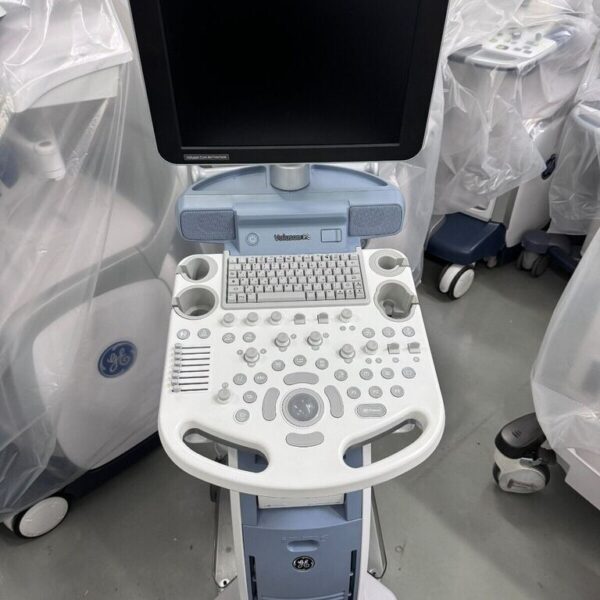
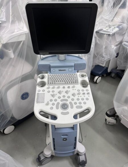
The GE Versana Premier ultrasound machine is a new color console offering from GE Healthcare with a 5 year warranty. This shared service system is designed for cardiac, point of care and women’s health imaging applications. The 21.5 inch monitor includes a fully articulating arm with a color touch screen and ergonomic keyboard, helping streamline your work. Multiple exam protocols and applications help you diagnose a wide spectrum of patient conditions. Intuitive automated tools streamline workflow. The system boots up quickly and operates quietly. It is compact to fit small spaces and easy to maneuver between rooms. The new GE Versana Premier ultrasound machine includes 4 hours of online training from GE, extensive training can be added. The system includes 4 probe ports, gel warmer and integrated speakers. Imaging modes include: Color Flow, PDI, Directional PDI, M Mode, Color M Mode, Pulse Wave Doppler. See below for additional software options you can add on to the system.Scan Assistant
Provide ultrasound sonographers with predefined standardized exam protocols that walk them through all steps of image acquisition, saving keystrokes, reducing stress and fatigue, and maximizing exam consistency.3D/4D
Visualize all three image planes within a scan volume in real time; especially useful in OB examinations. 3D image rendering and display provide fast reconstruction of anatomical sweeps. 4D provides continuous, high-volume acquisition of 3D images. 4D adds the dimension of movement to a 3D image.Whizz image tuning
Just touch the Whizz button on your console once. Without pressing it again, Whizz continuously and automatically optimizes the image, even as you move from one organ or structure to another.Effective exam follow-up
For follow-up exams, the Follow-up Tool automatically identifies the same ultrasound probe, preset, depth, gain and frequency settings as used in the prior scan. The current real-time ultrasound image displays side-by-side with the prior image for easy comparison.Insite Technology
GE advanced remote service technology and remote monitoring diagnostics and repair; instantly connect your ultrasound machine with a GE service expert to resolve many issues remotely on the spot. | SIUI CTS-800 Veterinary Ultrasound Machine with Linear Rectal Probe
This small and convenient “Palm-Sized” portable veterinary ultrasound scanner fits in the palm of your hand, and is mostly used for farm animals. Its long battery life and waterproof features makes it the perfect ultrasound scanner for veterinarians on the field.The screen of the SIUI CTS-800V will rotate horizontally or vertically, according to how you are holding it.Gravity Sensor: You can operate the ultrasound system either in transversal layout or vertical layout. It can automatically adapt to two different layouts when you rotate it.Grid for estimation: With grid on the image area you can easily estimate the size of the target substance like follicle fertilized egg etc.SPECIFICATIONS SIUI CTS-800 Veterinary Ultrasound Machine with Linear Rectal ProbeMonitor: 7-inch WVGA LCD monitorWeight: 0.8kg Environmental rating: IP54 (main unit) IP67 (probe head) Battery: 3 hours operation time Display mode: B 2B ZOOM B B/M M Depth: Max to 250mm B gain: 0-100 Image storage: 256 frames Cine loop: 256 frames Probe: L50/7.5 MHz linear rectal probe (standard) L65/5.0 MHz linear rectal probe (optional) R20/5.0 MHz micro convex probe (optional)
– Main unit with 7-inch LCD monitor.
– 1 Probe connector.
– 1 Li-ion battery. 3 hours operation time.
– 1 Adaptor.
– 1 Car Charger.
– Belt and sun shield.
– 1 USB port (Mini).
– Carrying case.PROBES– Rectal Linear L7FVC (6.5/7.1/7.5/8.3/9.0MHz).
– Rectal Linear L5FVC (4.0/4.7/5.0/5.5/6.2MHz).
– Backfat Linear L1FC (150mm, 2.5/3.0/3.5/4.0/5.0MHz).
– Convex C3FC (2.5/3.0/3.5/4.0/5.0MHz).
– Microconvex C5FC (3.5/4.0/5.0/6.5/7.1MHz).CTS-800 is a palm-sized veterinary ultrasound system. Features Weight: 0.8kg Environmental rating: IP54 (main unit) IP67 (probe head) Battery: 3 hours operation time Display mode: B 2B ZOOM B B/M M Depth: Max to 250mm B gain: 0-100 Image storage: 256 frames Cine loop: 256 frames Probe: L50/7.5 MHz linear rectal probe (standard) L65/5.0 MHz linear rectal probe (optional) R20/5.0 MHz micro convex probe (optional) | Toshiba Aplio 300 Ultrasound Machine
Toshiba Aplio 300 Ultrasound Machine Platinum and CV color Doppler ultrasound machines represent the more affordable versions of Canon’s high-end Aplio series of ultrasound machines. shared service machine or CV ultrasound includes Toshiba’s more advanced imaging innovations, including advanced compound imaging, speckle reduction, and tissue enhancement technologies. Additionally, the features 3D/4D imaging with high-density volume imaging.Toshiba Aplio 300 Ultrasound Machine For advanced performance, you can rely on day after day, look no further than . Cost-effective, productive and versatile, the system offers superior imaging with outstanding depth and detail for the widest range of clinical specialties. Lighter and with a smaller footprint, is designed to increase mobility, to optimize viewing, and to provide you with an ergonomic workplace. Products in general look like a technology oriented product, rather than a clinical oriented product. With this series, Canon has achieved fully digital beamforming technology, which is fully integrated into the Aplio Platinum series. The weight of each Platinum series is still high, above 320 lbs. The monitor size is 17-inches, but upgradable to 19-inches. Although the holds good quality and reputation in Radiology, they still need to be improved in cardiology and OB/GYN’s workflow, automation, and 4D quality, comparing GE or Samsung products to satisfy customers. - Shared Service
- Next Generation Precision Imaging
- ApliPure
- 3D/4D Imaging
- Advanced Dynamic Flow
- Contrast Harmonic Imaging
- Strain Elastography
- Gel Warmer
- Panoramic Option
- Stress Echo Option
- TDI Tissue Doppler Imaging
- HPRF High Pulsed Repetition Frequency
- DICOM Media Storage
- QuickScan
- 17″ LCD Monitor with 1280×1027 resolution and 170-degree viewing angle
- Trapezoidal Imaging
- TissuePure Speckle Reduction Imaging
- Compound Imaging
- IMT Intima-Media Thickness
- DICOM SR for Cardiac, Vascular, and OB/GYN
With the launch of the Toshiba Aplio 300 in 2011, the popularity with Toshiba is growing. Great for OB/Gyn to Cardiac, it offers the following:
• Differential Tissue Harmonics (D-THI): Enables uniformity in the near and far fields, especially while scanning at increased depth. • iStyleTM+ Productivity Suite: Delivers ergonomic relief by minimizing keystrokes and reducing exam times with the industry’s most customizable user interface and preset management tools.
• Ergonomics: Features a high-resolution LCD screen on a four-axis arm and handle, adjustable console, lighter weight and smaller footprint. | GE Voluson P8 Ultrasound MachineTHE VALUE OF ultrasound machine INNOVATIONAdvancing technology for effortless scanningSimply place the probe to get the information you need, without time-consuming modifications. Built Core Architecture, the Ultrasound Performance Series delivers excellent clinical images across all types of patients, quickly and easily.* Excellent clarity and detail in 2D images.
* Expand clinical confidence with 3D/4D technology.
* Superb sensitivity into anatomy and function with Advanced Color Doppler.
* Consistent imaging even in difficult-to-image patients with exceptional penetration.Obtain views not possible with 2D scanning with 3D imaging – offering another anatomical perspective for greater diagnostic certainty.Display more anatomical information in a single image with wide-sector abdominal and endocavitary probes.Optimize your skills by accessing educational videos directly on the ultrasound system.Explore 3D Printing for rapid clinical prototyping, and parent bonding.INSPIRING INTERACTIONS
Eliminate barriers while enhancing relationsDecrease complexity and increase exam consistency with Voluson’s Sono-automation technologies* Easily acquire surface rendered images with SonoRenderlive – Automates render line placement in 3D/4D imaging.
* Reduce keystrokes with SonoBiometry – semi-automated biometry measurements (BPD, HC, AC, FL and HL).Discover a sleek, lightweight system that fits into small spaces, enabling effortless scanning and reporting – A battery pack offers 20 minutes of scan time and ~15 second wake time from sleep mode.Protect sensitive data with Voluson ultrasound systems advanced security features including Whitelisting and HD encryption.Text or email images to patients or colleagues directly from the console, for an instant connection with TricefyTM inside.Count on responsive service and support from dedicated staff and programs that truly meet your training, equipment maintenance, transducer protection, and flexible financing. The GE Voluson P8 Ultrasound Machine is an excellent entry-point to the Voluson family and an affordable option to enjoy all benefits of the legacies it has built over decades. The Voluson P8 was developed based on the Voluson S6, S8, and E series and was introduced in the market in 2012. The imaging concept and design of the GE Voluson P8 is primarily focused and optimized for OB/GYN applications and is delivered through GE’s advanced imaging technologies like Coded Harmonic Imaging with Pulse Inversion technology, Speckle Reduction Imaging, and CrossXBeam (Compound Resolution Imaging). GE P8 Ultrasound Machine also provides an intuitive and easy-to-use keyboard from the Voluson family which allows for the immediate performance of everyday scanning.One of the major strengths of the GE P8 is the integration of several of GE’s unique automation technologies. Auto Optimization for 2D and Doppler helps enhance contrast resolutions at the touch of a button. Sono Biometry, SonoNT/IT, SonoAVCfollicle, SonoL&D, and Sono Render Start all improve the scanning and diagnosis efficiency of key exam operations by reducing the number of keystrokes needed and by enhancing the reproducibility of measurements. Application training GE Voluson P8 Ultrasound Machine
KPI’s on-staff sonographer can provide onsite applications training or remote training via video conference at a set price plus travel costs. A pre-recorded video training course is included in the sale, lease or rental of the Voluson P8GE Voluson P8 Dimensions & Weight
Width: 420 mm (16.5 in)
Depth: 670 mm (26.4 in)
Height: 1495 mm (59 in)
Weight: 143 lbs/65 kgGE Voluson P8 Specifications:
System processing channel counts up to 161,616 by configuration
Minimum Depth of Field: 0 – 1 cm (Zoom, probe dependent)
Maximum Depth of Field: 0 – 30 cm (probe dependent)
Continuous dynamic receive focus/continuous dynamic receive aperture
Adjustable dynamic range up to 255 dBGE Voluson P8 Electrical power:
Nominal input voltage: 100-240 VAC, frequency 50/60 HzReview - GE Voluson P8 Ultrasound Machine The GE Voluson P8 is an excellent entry-point to the Voluson family and an affordable option to enjoy all benefits of the legacies it has built over decades. The Voluson P8 was developed based on the Voluson S6, S8, and E series and was introduced in the market in 2012. The imaging concept and design of the GE Voluson P8 is primarily focused and optimized for OB/GYN applications and is delivered through GE’s advanced imaging technologies like Coded Harmonic Imaging with Pulse Inversion technology, Speckle Reduction Imaging, and CrossXBeam (Compound Resolution Imaging). The GE Voluson P8 also provides an intuitive and easy-to-use keyboard from the Voluson family which allows for the immediate performance of everyday scanning.One of the major strengths of the GE Voluson GE Voluson P8 Ultrasound MachineV is the integration of several of GE’s unique automation technologies. Auto Optimization for 2D and Doppler helps enhance contrast resolutions at the touch of a button. SonoBiometry, SonoNT/IT, SonoAVCfollicle, SonoL&D, and SonoRenderStart all improve the scanning and diagnosis efficiency of key exam operations by reducing the number of keystrokes needed and by enhancing the reproducibility of measurements.3D HDlive is one of the primary features that sets the GE Voluson P8 apart from its competitors, namely the UGEO H60 and SonoAce R7 from Samsung, and the Philips ClearVue 650. Investors will find this high-resolution 3D imaging technology cost-efficient with added value for physicians and patients. GE Voluson P8 probes / transducers
Convex Probe
4C-RSLinear Probe
12L-RSEndocavitary Probe
E8C-RSPhased Array Probe
3Sc-RS3D/4D Convex Probe
RAB2-6-RS3D/4D Endocavitary Probe
RIC5-9W-RSGE Voluson P8 Features:
• 17” High Resolution LCD LED Flat Panel Display
• 3 Active Probe Ports
• Integrated HD (500 GB)
• 3 USB Ports for External Peripherals
• 2 USB Ports for On-board Peripherals
• AO(Automatic Optimization)
• CrossXBeam(Spatial Compounding Imaging)
• SRI(Speckle Reduction Imaging)
• B-Steer
• Coded Phased Inversion Harmonic Imaging
• HD-flow
• 3/4D(Real time)
• Virtual Convex
• Patient Information Database
• SonoBiometry
• SonoRender Start
• Raw Data fileMeasurement & Calculations including worksheets/report for:
• OB Measurement, Calculations & Reports
• GYN Measurement, Calculations & Reports
• Vascular Measurement, Calculations & Reports
• Cardio Measurement, Calculations & Reports
• Abdominal Measurement, Calculations & Reports
• Small-Parts Measurement, Calculations & Reports
• Urology Measurement, Calculations & Reports
• Pediatrics Measurement, Calculations & Reports
• Musculoskeletal Measurement, Calculations & Reports
• Neurology Measurement, Calculations & ReportsOptions : GE Voluson P8 Ultrasound Machine
• Sony Digital UP-D711 Termal Printer
• Sony Fixture Kit for Digital UP-D711 Thermal Printer
• Sony Digital UP-D25 Color Thermal Printer
• Sony Digital UP-D897 BW Thermal Printer
• Mitsubishi P93W/E Thermal Printer
• Mitsubishi P95DW Thermal Printer
• Footswitch MKF2-MED USB GP26
• USB ECG Kits (AHA/IEC)Supplies:
• Aquasonic ultrasound gel
• Sono ultrasound wipes
• Sony UPP-110HG thermal printing paper
• Sony UPC-21L color thermal printing pack
• Mitsubishi KP95HG thermal roll paper (new)
• Mitsubishi KP65HM-CE High density thermal paperports:
• 3 USB Ports for External Peripherals
• 2 USB Ports for On-board Peripherals
• RJ45 LAN Port
• 1 HDMI Out Port
• 1 AUDIO Out Portimage storage:
• Storage formats– DICOM files(Single- or Multi-frame) DCM, DICOM Files with DICOMDIR
– Raw Data file (Proprietary format)
– Export: BMP, TIFF, JPEG, MPEG 4, MS Video 1• Storage Devices: USB Memory Stick
• DVD-RW Storage
• HDD Image StorageOptions
• Static 3D Mode
• 3/4D Advanced
• HD Live(3D Only)
• SonoNT/SonoIT
• SonoL&D
• 4D View PC Software
• DICOM 3
• Extended View(XTD)
• Anatomical M-mode
• Report EditorApplications
• Abdominal
• Gynecology
• Small parts
• Vascular
• Pediatrics
• Cardiology
• Urology
• Musculoskeletal(MSK)
• Breast | Samsung Medison SonoAce R3 Ultrasound Machine
SonoAce R3 is a portable color ultrasound system with comprehensive diagnostic capabilities. Full Spectrum Imaging™ (FSI™) combines with Speckle Reduction Filter™ (SRF™) to deliver superior quality images, even in the challenging circumstances. The compact and user-friendly design features of the SonoAce R3 ensure portability and manoeuvrability in a lightweight unit. Sporting a 15-inch LCD monitor with Full-Featured Workflow, the SonoAceR3 offers your organisation flexibility, without compromising on precision and efficiency.Slim and Compact DesignThe compact design of SonoAce R3 offers Various-Featured Workflow with a thumbnail menu and shortcut, together with functional key groupings for increased efficiency. The user-friendly design combines a wide-view 15-inch LCD monitor with backlit control keys, customisable measurement package and body markers. The SonoAce R3 features 2 probe ports, 3 USB ports, rapid boot up capability and DICOM 3 compatible image filing. This slim, lightweight unit also has a convenient carrying handle for portability and flexibility.Samsung Medison SonoAce R3 Ultrasound MachineFull Spectrum Imaging™ (FSI™) produces the penetration capabilities associated with the lower frequencies, while still maintaining the fine pixel uniformity associated with the higher frequencies. This combination consistently delivers superior quality images, even in the challenging diagnostic circumstances. The resulting clarity delivers improvements in accuracy and efficiency for greater confidence in your diagnostic procedures. Harmonic Imaging is a technique that records higher frequency information, resulting in greater resolution and fewer artifacts.Synthetic Aperture
The physical radio channel may present some limits to imaging which can be overcome by using Synthetic Aperture Control software. This method uses information from diverse sources to form a more better picture of the area under examination. It creates one single scan line by performing the TX and RX twice to create enhanced penetration and resolution. Synthetic Aperture Control technology delivers a comprehensive picture and to aid accurate diagnosis.QuickScan
QuickScan has been designed to increase workflow efficiency by automatically optimising key imaging parameters at the touch of a button. The resulting increase in efficiency allows your organisation to offer patients an improved service with faster and more accurate diagnoses. This reliable and safe system helps your clinic or hospital to deliver a superior patient experience.Features
Full Spectrum ImagingTM
Quick ScanTM
Color and Power Doppler
Harmonic imaging
Freehand 3D
3D Multi Planar Imaging
Sonoview Image Management
PW Doppler
15″ LCD monitor
Two probe ports
Lightweight | Siemens Acuson Sequoia 512 LCD Ultrasound Machine18″ Flat panel LCD Monitor–Progressive Media & Display II Package, Cardiac/OB/Vascular Calc. Package, Signature II Option, AUX, CPS Cardiology, CPS General Cadene Trigg Burst, Calc Data to MO, Card Cadence II Contrast Quant, Cornoary Flow reserve, Dicome Store/Print, DTI (Doppler Tissue Imaging), High HR ECG, MIS/MIF, Modality Worklist Native TEQ, Native TEQ, NTHI (Native Tissue Harmonics), Spectral TEQ, TEQ (Tissue Equalization), VVIFeatures- Shared Service
- Native Patient Specific Imaging
- Four Sight TEE view
- Axius quantitative strain rate imaging
- Cadence contrast pulse sequencing technology
- Native Tissue Harmonic Imaging
- Native TEQ ultrasound technology
- Color doppler, Color angio and b-color
- PW/CW Doppler
- M-Mode
- Folding Flat Panel Display
- DICOM capabilities
Applications- Abdominal
- Breast
- OB/GYN
- Vascular
- Pediatrics
- Cardiology
- Musculoskeletal
- Neonatal
Acuson Sequoia 512 Features:- Imaging: Gray-scale 2-D, M-mode, SST Color Doppler, Solo Spectral Doppler, 3-D Surface Rendering, Native Tissue Harmonic Imaging, Auto Doppler, M-Mode, PW Doppler, Color Doppler, Color Power Doppler
- 19" color CRT monitor
- Native tissue harmonic imaging (NTHI)
- DIMAQ: image management
- Advanced vascular analysis
- Advanced spatial compounding
- Native patient specific imaging
- Mitral valve assessment
- CPS contrast pulse sequencing technology
- Clarifying vascular enhancement technology
- High resolution color flow
- Vascular, and General Imaging calculation packages
- Coherent beamformer technology/spatial compounding
- Space/time resolution control
- Cine and zoom/RES
Compatible Ultrasound Probes and Transducers:- Acuson V5Ms TEE Probe
- Acuson 4C1 Curved Array Probe
- Acuson 6C2 Curved Array Probe
- Acuson 8C4 Curved Array Probe
- Acuson 4V1 Vector Array Probe
- Acuson EV-8C4 Endocavitary Probe
- Acuson EC-10C5 Endocavitary Probe
- Acuson 6L3 Linear Array Probe
- Acuson 8L5T Intraoperative Probe
- Acuson 8L5 Linear Array Probe
- Acuson 9L4 Linear Array Probe
- Acuson 15L8 Linear Array Probe
- Acuson 17L5 Linear Array Probe
The Sequoia 512 with LCD ultrasound machine uniquely minimizes automatic image optimization time by up to 80% to make way for an easier and speedier ultrasound test time as well as allow for a more confident diagnosis each time. As a patient-specific imaging ultrasound, it has a unique matched response capacity for accurate image quality, sharp clinical specificity, and optimized diagnostic outcomes that makes infinitesimal pathologies and other anatomical detail in the most difficult-to-image patients becomes more vivid and visible. |





Reviews
There are no reviews yet.