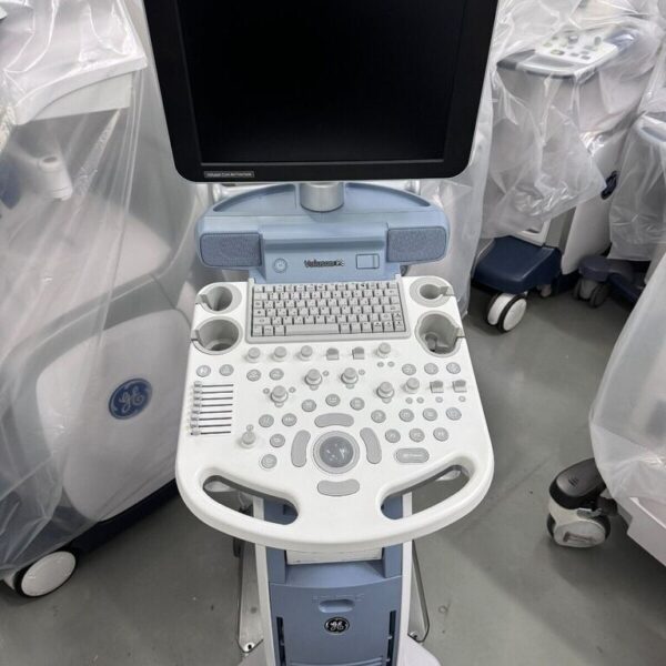
| Content | GE Versana Premier Ultrasound Machine
The GE Versana Premier ultrasound machine is a new color console offering from GE Healthcare with a 5 year warranty. This shared service system is designed for cardiac, point of care and women’s health imaging applications. The 21.5 inch monitor includes a fully articulating arm with a color touch screen and ergonomic keyboard, helping streamline your work. Multiple exam protocols and applications help you diagnose a wide spectrum of patient conditions. Intuitive automated tools streamline workflow. The system boots up quickly and operates quietly. It is compact to fit small spaces and easy to maneuver between rooms. The new GE Versana Premier ultrasound machine includes 4 hours of online training from GE, extensive training can be added. The system includes 4 probe ports, gel warmer and integrated speakers. Imaging modes include: Color Flow, PDI, Directional PDI, M Mode, Color M Mode, Pulse Wave Doppler. See below for additional software options you can add on to the system.Scan Assistant
Provide ultrasound sonographers with predefined standardized exam protocols that walk them through all steps of image acquisition, saving keystrokes, reducing stress and fatigue, and maximizing exam consistency.3D/4D
Visualize all three image planes within a scan volume in real time; especially useful in OB examinations. 3D image rendering and display provide fast reconstruction of anatomical sweeps. 4D provides continuous, high-volume acquisition of 3D images. 4D adds the dimension of movement to a 3D image.Whizz image tuning
Just touch the Whizz button on your console once. Without pressing it again, Whizz continuously and automatically optimizes the image, even as you move from one organ or structure to another.Effective exam follow-up
For follow-up exams, the Follow-up Tool automatically identifies the same ultrasound probe, preset, depth, gain and frequency settings as used in the prior scan. The current real-time ultrasound image displays side-by-side with the prior image for easy comparison.Insite Technology
GE advanced remote service technology and remote monitoring diagnostics and repair; instantly connect your ultrasound machine with a GE service expert to resolve many issues remotely on the spot. | GE Vscan Access Portable Ultrasound Device
With Vscan Access, midwives, paramedics, clinical officers, and general practitioners have the means to:Conduct examinations that may result in the early detection of potentially life-threatening pregnancy complications
Estimate gestational age and delivery date to help better plan and manage deliveries compared to manual exam methods
Guide the critical “keep or refer” decisions over the course of antenatal visits
Wirelessly transfer data to patients, referral facilities or remote expertFor more information or a quote on the GE Healthcare Vscan Access Portable Ultrasound machine; please contact us https://www.metrodentalmedicalcenter.com email enquiry for a direct reply.GE Vscan Access Portable Ultrasound DeviceUltrasound designed for primary health workers in developing nations to help assess pregnancy risks
Maternal and newborn health remains one of our most pressing priorities. Despite advances, many countries need significant investment and cross-sector collaboration to help reduce pregnancy-related mortality. GE’s latest innovation is an ultrasound device designed for primary health workers to assess pregnancy risks early, and help expand the reach of quality care to mothers who need it most. The World Health Organization (WHO) estimates that “70% of medical equipment coming from the most developed nations does not work in developing world facilities.” To avoid these pitfalls, GE sought to re-think ultrasound from the ground-up. An global team of engineers, designers and health experts, guided by a Human-Centred Design process worked over 3 years to make VScan Access: SIMPLE for primary health workers to learn and use effectively DURABLE to last for years in tough* environments INNOVATIVE to integrate mHealth solutions AFFORDABLE to enable national scale-upThe Vscan Access developed by GE Healthcare is a new portable ultrasound system designed specifically for primary healthcare workers—such as midwives, general practitioners, paramedics and clinical officers—in low-resource settings.EvaluationTraditionally, manufacturers have vied to increase the resolution and performance of ultrasonic diagnostic equipment, and such equipment has gotten more expensive and grown larger in size. With this model, the manufacturer effectively narrowed down its functionality and downsized its main unit based on an accurate assessment of medical care needs. This has also raised applicability in medical care scenes due to its improved portability, making it the successor to a medical device that was highly praised in the past. Without excessively increasing the size of its main unit, which was characteristic of previous models, its display was moderately enlarged, which is among its features that improve its usability. This is an outstanding product that accurately reflects the needs found at the front lines of medicine. | SonoSite X Porte Ultrasound Machine
SonoSite X Porte Ultrasound Machine represents an entirely new approach to clinical ultrasound. Its imaging, features and educational resources are fluidly brought together in a convenient, all-in-one kiosk design. At the sweep of your hand, it responds so quickly and intelligently to your imaging needs, you’ll know it was created precisely for professionals like you. Its self-explanatory control panel makes system navigation a breeze, and its sealed touch screen has no buttons for pathogens to hide behind.
X-Porte’s slender profile makes it easy to maneuver alongside beds and exam tables for point-of-care visualization and procedures. For portability and durability during transport, its screen folds down and its stand lowers making X-Porte even more compact for navigating busy corridors. The X-Porte ultrasound core can be detached easily from the kiosk to provide another configuration option. For servicing, nothing could be more convenient than X-Porte’s five-year warranty and removable engine.SonoSite X Porte Ultrasound Machine Benefits:
– Large monitor tilts and turns for optimal viewing in a crowded room or for interacting with patients
– With no buttons, the control panel and monitor may be cleaned and disinfected with ease
– Can be operated with gloves and sterile drape
– High-definition imaging for improved decision making confidence
– Streamline workflow with remote worksheets, barcode scanning, and menu customization
– Real-time learning with 81 onboard tutorials to view instructional videos while performing live scans
– Modular design and removable engine for easy servicing of components for decreased downtimeSonoSite X Porte Ultrasound Machine SYSTEM SPECIFICATIONS:
Stand Dimensions : 26.4″ L x 21.2″ W
Stand Height (max) : 65″ (monitor up)
Stand Height (min) : 42.2″ (monitor down)
Height Adjustment : 9″ travel
HD Monitor : 19″ diagonally (minimum)
Control Panel : 12.1″ capacitive touchscreen
System Controls : Multi-touch gestures
Tilt Adjustment : 0 to 110 degrees
Clinical Monitor Viewing : 85 degrees left and right, 85 degrees up, 85 degrees down
Architecture : All digital broadband
Dynamic Range : Up to 183 dB
Gray Scale : 256 shades
HIPAA Compliance : Comprehensive toolsetIMAGE MODES
2D Broadband Imaging
Tissue Harmonic Imaging
Pulse Inversion Harmonic Imaging
M-Mode
Velocity Color Doppler
Color Power Doppler
Pulsed Wave Doppler (update and simultaneous)
Pulsed Wave Tissue Doppler
Continuous Wave Doppler, ECGIMAGE PROCESSING
Extreme definition imaging technology
SonoAdapt Tissue Optimization
SonoHD2 Imaging Technology
SonoMB Multibeam Technology
Dual Imaging
Dual Color Imaging
AutoGain and AutoGain Brightness Adjust
Restore Default Gains
Dynamic Range
Duplex Imaging
8x Zoom Capability
Cine Processing: Dynamic Range, Zoom
2D Image Optimization: Res, Gen, Pen
Overall Gain, Near and Far Field Gain Control
Color and Doppler Flow Optimization (low, medium, high)
Color Variance Mode
2D Reduced Imaging Sector (steerable)TRANSDUCERS:
– C11xp 8-5 MHz Curved
Exam Types: Abdominal, Arterial, Neonatal, Nerve, Venous
Scan depth: 15 cm– C35xp 8-3 MHz Curved
Exam Types: Abdominal, MSK, Nerve
Scan depth: 16 cm– C60xp 5-2 MHz Curved
Exam Types: Abdominal, Gynecology, MSK, Nerve, Obstetrics
Scan depth: 30 cm– ICTxp 9-5 MHz Curved
Exam Types: Gynecology, Obstetrics
Scan depth: 15 cm– HFL38xp 13-6 MHz Linear
Exam Types: Arterial, Breast, Lung, MSK, Nerve, Small Parts, Venous
Scan depth: 6 cm– HFL50xp 15-6 MHz Linear
Exam Types: Breast, MSK, Nerve, Small Parts
Scan depth: 6 cm– HSL25xp 13-6 MHz Linear
Exam Types: Arterial, Lung, MSK, Nerve, Ophthalmic, Superficial, Venous
Scan depth: 6 cm– L25xp 13-6 MHz Linear
Exam Types: Arterial, Lung, MSK, Nerve, Ophthalmic, Superficial, Venous
Scan depth: 6 cm– L38xp 10-5 MHz Linear
Exam Types: Arterial, Lung, Nerve, Small Parts, Venous
Scan depth: 9 cm– P10xp 8-4 MHz Phased
Exam Types: Abdominal, Cardiac, Neonatal
Scan depth: 15 cm– rP19xp 5-1 MHz Phased
Exam Types: Abdomen, Cardiac, Lung, Obstetrics, Orbital, Transcranial
Scan depth: 35 cm– TEExp 8-3 MHz Phased
Exam Types: Cardiac
Scan depth: 18 cm– D2xp 2MHz Pencil
Exam Types: Cardiac
Scan depth: N/A | Siemens Acuson Sequoia 512 LCD Ultrasound Machine18″ Flat panel LCD Monitor–Progressive Media & Display II Package, Cardiac/OB/Vascular Calc. Package, Signature II Option, AUX, CPS Cardiology, CPS General Cadene Trigg Burst, Calc Data to MO, Card Cadence II Contrast Quant, Cornoary Flow reserve, Dicome Store/Print, DTI (Doppler Tissue Imaging), High HR ECG, MIS/MIF, Modality Worklist Native TEQ, Native TEQ, NTHI (Native Tissue Harmonics), Spectral TEQ, TEQ (Tissue Equalization), VVIFeatures- Shared Service
- Native Patient Specific Imaging
- Four Sight TEE view
- Axius quantitative strain rate imaging
- Cadence contrast pulse sequencing technology
- Native Tissue Harmonic Imaging
- Native TEQ ultrasound technology
- Color doppler, Color angio and b-color
- PW/CW Doppler
- M-Mode
- Folding Flat Panel Display
- DICOM capabilities
Applications- Abdominal
- Breast
- OB/GYN
- Vascular
- Pediatrics
- Cardiology
- Musculoskeletal
- Neonatal
Acuson Sequoia 512 Features:- Imaging: Gray-scale 2-D, M-mode, SST Color Doppler, Solo Spectral Doppler, 3-D Surface Rendering, Native Tissue Harmonic Imaging, Auto Doppler, M-Mode, PW Doppler, Color Doppler, Color Power Doppler
- 19" color CRT monitor
- Native tissue harmonic imaging (NTHI)
- DIMAQ: image management
- Advanced vascular analysis
- Advanced spatial compounding
- Native patient specific imaging
- Mitral valve assessment
- CPS contrast pulse sequencing technology
- Clarifying vascular enhancement technology
- High resolution color flow
- Vascular, and General Imaging calculation packages
- Coherent beamformer technology/spatial compounding
- Space/time resolution control
- Cine and zoom/RES
Compatible Ultrasound Probes and Transducers:- Acuson V5Ms TEE Probe
- Acuson 4C1 Curved Array Probe
- Acuson 6C2 Curved Array Probe
- Acuson 8C4 Curved Array Probe
- Acuson 4V1 Vector Array Probe
- Acuson EV-8C4 Endocavitary Probe
- Acuson EC-10C5 Endocavitary Probe
- Acuson 6L3 Linear Array Probe
- Acuson 8L5T Intraoperative Probe
- Acuson 8L5 Linear Array Probe
- Acuson 9L4 Linear Array Probe
- Acuson 15L8 Linear Array Probe
- Acuson 17L5 Linear Array Probe
The Sequoia 512 with LCD ultrasound machine uniquely minimizes automatic image optimization time by up to 80% to make way for an easier and speedier ultrasound test time as well as allow for a more confident diagnosis each time. As a patient-specific imaging ultrasound, it has a unique matched response capacity for accurate image quality, sharp clinical specificity, and optimized diagnostic outcomes that makes infinitesimal pathologies and other anatomical detail in the most difficult-to-image patients becomes more vivid and visible. | Samsung Medison SonoAce R3 Ultrasound Machine
SonoAce R3 is a portable color ultrasound system with comprehensive diagnostic capabilities. Full Spectrum Imaging™ (FSI™) combines with Speckle Reduction Filter™ (SRF™) to deliver superior quality images, even in the challenging circumstances. The compact and user-friendly design features of the SonoAce R3 ensure portability and manoeuvrability in a lightweight unit. Sporting a 15-inch LCD monitor with Full-Featured Workflow, the SonoAceR3 offers your organisation flexibility, without compromising on precision and efficiency.Slim and Compact DesignThe compact design of SonoAce R3 offers Various-Featured Workflow with a thumbnail menu and shortcut, together with functional key groupings for increased efficiency. The user-friendly design combines a wide-view 15-inch LCD monitor with backlit control keys, customisable measurement package and body markers. The SonoAce R3 features 2 probe ports, 3 USB ports, rapid boot up capability and DICOM 3 compatible image filing. This slim, lightweight unit also has a convenient carrying handle for portability and flexibility.Samsung Medison SonoAce R3 Ultrasound MachineFull Spectrum Imaging™ (FSI™) produces the penetration capabilities associated with the lower frequencies, while still maintaining the fine pixel uniformity associated with the higher frequencies. This combination consistently delivers superior quality images, even in the challenging diagnostic circumstances. The resulting clarity delivers improvements in accuracy and efficiency for greater confidence in your diagnostic procedures. Harmonic Imaging is a technique that records higher frequency information, resulting in greater resolution and fewer artifacts.Synthetic Aperture
The physical radio channel may present some limits to imaging which can be overcome by using Synthetic Aperture Control software. This method uses information from diverse sources to form a more better picture of the area under examination. It creates one single scan line by performing the TX and RX twice to create enhanced penetration and resolution. Synthetic Aperture Control technology delivers a comprehensive picture and to aid accurate diagnosis.QuickScan
QuickScan has been designed to increase workflow efficiency by automatically optimising key imaging parameters at the touch of a button. The resulting increase in efficiency allows your organisation to offer patients an improved service with faster and more accurate diagnoses. This reliable and safe system helps your clinic or hospital to deliver a superior patient experience.Features
Full Spectrum ImagingTM
Quick ScanTM
Color and Power Doppler
Harmonic imaging
Freehand 3D
3D Multi Planar Imaging
Sonoview Image Management
PW Doppler
15″ LCD monitor
Two probe ports
Lightweight | GE Voluson P8 Ultrasound MachineTHE VALUE OF ultrasound machine INNOVATIONAdvancing technology for effortless scanningSimply place the probe to get the information you need, without time-consuming modifications. Built Core Architecture, the Ultrasound Performance Series delivers excellent clinical images across all types of patients, quickly and easily.* Excellent clarity and detail in 2D images.
* Expand clinical confidence with 3D/4D technology.
* Superb sensitivity into anatomy and function with Advanced Color Doppler.
* Consistent imaging even in difficult-to-image patients with exceptional penetration.Obtain views not possible with 2D scanning with 3D imaging – offering another anatomical perspective for greater diagnostic certainty.Display more anatomical information in a single image with wide-sector abdominal and endocavitary probes.Optimize your skills by accessing educational videos directly on the ultrasound system.Explore 3D Printing for rapid clinical prototyping, and parent bonding.INSPIRING INTERACTIONS
Eliminate barriers while enhancing relationsDecrease complexity and increase exam consistency with Voluson’s Sono-automation technologies* Easily acquire surface rendered images with SonoRenderlive – Automates render line placement in 3D/4D imaging.
* Reduce keystrokes with SonoBiometry – semi-automated biometry measurements (BPD, HC, AC, FL and HL).Discover a sleek, lightweight system that fits into small spaces, enabling effortless scanning and reporting – A battery pack offers 20 minutes of scan time and ~15 second wake time from sleep mode.Protect sensitive data with Voluson ultrasound systems advanced security features including Whitelisting and HD encryption.Text or email images to patients or colleagues directly from the console, for an instant connection with TricefyTM inside.Count on responsive service and support from dedicated staff and programs that truly meet your training, equipment maintenance, transducer protection, and flexible financing. The GE Voluson P8 Ultrasound Machine is an excellent entry-point to the Voluson family and an affordable option to enjoy all benefits of the legacies it has built over decades. The Voluson P8 was developed based on the Voluson S6, S8, and E series and was introduced in the market in 2012. The imaging concept and design of the GE Voluson P8 is primarily focused and optimized for OB/GYN applications and is delivered through GE’s advanced imaging technologies like Coded Harmonic Imaging with Pulse Inversion technology, Speckle Reduction Imaging, and CrossXBeam (Compound Resolution Imaging). GE P8 Ultrasound Machine also provides an intuitive and easy-to-use keyboard from the Voluson family which allows for the immediate performance of everyday scanning.One of the major strengths of the GE P8 is the integration of several of GE’s unique automation technologies. Auto Optimization for 2D and Doppler helps enhance contrast resolutions at the touch of a button. Sono Biometry, SonoNT/IT, SonoAVCfollicle, SonoL&D, and Sono Render Start all improve the scanning and diagnosis efficiency of key exam operations by reducing the number of keystrokes needed and by enhancing the reproducibility of measurements. Application training GE Voluson P8 Ultrasound Machine
KPI’s on-staff sonographer can provide onsite applications training or remote training via video conference at a set price plus travel costs. A pre-recorded video training course is included in the sale, lease or rental of the Voluson P8GE Voluson P8 Dimensions & Weight
Width: 420 mm (16.5 in)
Depth: 670 mm (26.4 in)
Height: 1495 mm (59 in)
Weight: 143 lbs/65 kgGE Voluson P8 Specifications:
System processing channel counts up to 161,616 by configuration
Minimum Depth of Field: 0 – 1 cm (Zoom, probe dependent)
Maximum Depth of Field: 0 – 30 cm (probe dependent)
Continuous dynamic receive focus/continuous dynamic receive aperture
Adjustable dynamic range up to 255 dBGE Voluson P8 Electrical power:
Nominal input voltage: 100-240 VAC, frequency 50/60 HzReview - GE Voluson P8 Ultrasound Machine The GE Voluson P8 is an excellent entry-point to the Voluson family and an affordable option to enjoy all benefits of the legacies it has built over decades. The Voluson P8 was developed based on the Voluson S6, S8, and E series and was introduced in the market in 2012. The imaging concept and design of the GE Voluson P8 is primarily focused and optimized for OB/GYN applications and is delivered through GE’s advanced imaging technologies like Coded Harmonic Imaging with Pulse Inversion technology, Speckle Reduction Imaging, and CrossXBeam (Compound Resolution Imaging). The GE Voluson P8 also provides an intuitive and easy-to-use keyboard from the Voluson family which allows for the immediate performance of everyday scanning.One of the major strengths of the GE Voluson GE Voluson P8 Ultrasound MachineV is the integration of several of GE’s unique automation technologies. Auto Optimization for 2D and Doppler helps enhance contrast resolutions at the touch of a button. SonoBiometry, SonoNT/IT, SonoAVCfollicle, SonoL&D, and SonoRenderStart all improve the scanning and diagnosis efficiency of key exam operations by reducing the number of keystrokes needed and by enhancing the reproducibility of measurements.3D HDlive is one of the primary features that sets the GE Voluson P8 apart from its competitors, namely the UGEO H60 and SonoAce R7 from Samsung, and the Philips ClearVue 650. Investors will find this high-resolution 3D imaging technology cost-efficient with added value for physicians and patients. GE Voluson P8 probes / transducers
Convex Probe
4C-RSLinear Probe
12L-RSEndocavitary Probe
E8C-RSPhased Array Probe
3Sc-RS3D/4D Convex Probe
RAB2-6-RS3D/4D Endocavitary Probe
RIC5-9W-RSGE Voluson P8 Features:
• 17” High Resolution LCD LED Flat Panel Display
• 3 Active Probe Ports
• Integrated HD (500 GB)
• 3 USB Ports for External Peripherals
• 2 USB Ports for On-board Peripherals
• AO(Automatic Optimization)
• CrossXBeam(Spatial Compounding Imaging)
• SRI(Speckle Reduction Imaging)
• B-Steer
• Coded Phased Inversion Harmonic Imaging
• HD-flow
• 3/4D(Real time)
• Virtual Convex
• Patient Information Database
• SonoBiometry
• SonoRender Start
• Raw Data fileMeasurement & Calculations including worksheets/report for:
• OB Measurement, Calculations & Reports
• GYN Measurement, Calculations & Reports
• Vascular Measurement, Calculations & Reports
• Cardio Measurement, Calculations & Reports
• Abdominal Measurement, Calculations & Reports
• Small-Parts Measurement, Calculations & Reports
• Urology Measurement, Calculations & Reports
• Pediatrics Measurement, Calculations & Reports
• Musculoskeletal Measurement, Calculations & Reports
• Neurology Measurement, Calculations & ReportsOptions : GE Voluson P8 Ultrasound Machine
• Sony Digital UP-D711 Termal Printer
• Sony Fixture Kit for Digital UP-D711 Thermal Printer
• Sony Digital UP-D25 Color Thermal Printer
• Sony Digital UP-D897 BW Thermal Printer
• Mitsubishi P93W/E Thermal Printer
• Mitsubishi P95DW Thermal Printer
• Footswitch MKF2-MED USB GP26
• USB ECG Kits (AHA/IEC)Supplies:
• Aquasonic ultrasound gel
• Sono ultrasound wipes
• Sony UPP-110HG thermal printing paper
• Sony UPC-21L color thermal printing pack
• Mitsubishi KP95HG thermal roll paper (new)
• Mitsubishi KP65HM-CE High density thermal paperports:
• 3 USB Ports for External Peripherals
• 2 USB Ports for On-board Peripherals
• RJ45 LAN Port
• 1 HDMI Out Port
• 1 AUDIO Out Portimage storage:
• Storage formats– DICOM files(Single- or Multi-frame) DCM, DICOM Files with DICOMDIR
– Raw Data file (Proprietary format)
– Export: BMP, TIFF, JPEG, MPEG 4, MS Video 1• Storage Devices: USB Memory Stick
• DVD-RW Storage
• HDD Image StorageOptions
• Static 3D Mode
• 3/4D Advanced
• HD Live(3D Only)
• SonoNT/SonoIT
• SonoL&D
• 4D View PC Software
• DICOM 3
• Extended View(XTD)
• Anatomical M-mode
• Report EditorApplications
• Abdominal
• Gynecology
• Small parts
• Vascular
• Pediatrics
• Cardiology
• Urology
• Musculoskeletal(MSK)
• Breast |




Reviews
There are no reviews yet.