| Description | Confidently measure the human eye with a sophisticated cutting edge technology designed to enhance your ability to diagnose ocular errors faster, easier, and accurately. Ezers ERK-7800 is brilliantly designed technology measures the eye with ultra precision and speed to provide reliable data as the starting point of your refraction and contact lens fitting. It’s compact and modern design enables seamless operation and an enhanced patient comfort, making it the ideal choice in for any ophthalmic practice. Backed by an industry leading 2-years warranty! | Experience 6 analyses in 1 device: refraction, keratometry, topography, tonometry, pachymetry and dry eye analysis. | workflow, because some letters inspire confidence.With Neksia, Essilor combines years of experience in high-performance and precision edging into one of the most user-friendly and productive systems in the world. | The OPD-Scan III is an automated 5 in 1 instrument that combines topography, wavefront, autorefraction, keratometry, and pupillometry — allowing accurate and reliable analysis of corneal aberration. | The impression scan can be applied directly in 3Shape DentalDesigner
like any gypsum model scan and sets new standards for system
applicability. In combination with DentalDesigner the new D500
scanner brings unprecedented scan-to completed design time to the
market for digital dentistry. attractive opportunities in terms of
manufacturing cost, model creation speed and quality. | Acuson Cypress cardiovascular system PLUS delivers advanced ergonomics and superior performance in a highly portable package. TheCypress System PLUSis a highly miniaturized, all digital, phased array echocardiography system that provides complete studies and outstanding images – even on patients who are difficult to image. |
| Content | Ezer ERK-9000 Ezer Autorefractor KeratometerThe ERK-9000 offers your staff the opportunity to gather a wealth of precise information about the patient in one fast and easy test, making the remainder of your exam swift, simple and exact. In addition to the wealth of functions you’d expect from a top-of-the-line autorefractor/keratometer, the ERK-9000 features auto-shoot programming, an illumination mode, IOL mode and so much more.
Features:– Auto Refractor/Keratometer
– Auto-Shoot Programming
– Simple and Convenient User Environment
– IOL Mode
– Illumination Mode
– Easy Network Capability
– Wide Dioptric Measurement Range
– Accurate Iris and Pupil Measurement
– Ergonomic Joystick
– Motorized Chin Rest
– High-Speed Printer with Auto Paper Cut
– 2-Year WarrantyIncludes:Ezer ERK-9000 Ezer Autorefractor Keratometer– Power Supply Cable
– Model Eye ( S-5.0D or 5.12D or 5.25D, VD=12mm, R–7.95 )
– 2 rolls Printing Paper( Thermal Paper, Size : 57 X 50 mm )
– Dust cover ( Vinyl Cover: Use for long-term storage to prevent dust pollution )
– Operation ManualThe ERK-9000 combines all of the necessary visual pre-testing functions in one smartly designed instrument. Operators can perform refractometry and keratometry alone or simultaneously. The ERK-9000 is also quick. In fact, not even the unwanted effects of patient accommodation will stand in your way thanks to the unit’s valuable built-in auto-fogging mechanism.Auto-Shoot Programming
Users can program the ERK-9000 to shoot at their preferred mode. One click on the joystick button can take a single measurement, three continuous measurements or five continuous measurements.Simple and Convenient User Environment
The ERK-9000 features a user-friendly dashboard view. Sharp images with easy-to-understand icons make this 7.0-inch color TFT color touch screen LCD interface enjoyable to work with.
IOLMode
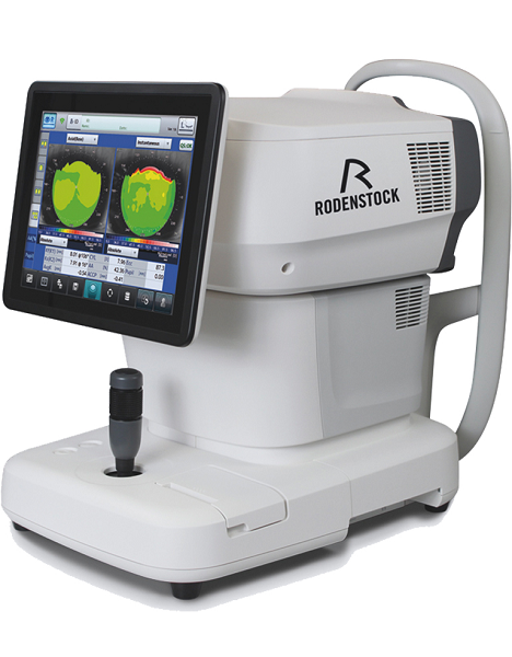
Patients presenting with cataract or IOL won’t present a challenge with the ERK-7800. In fact, now you can gather a wealth of information about natural lens opacity as well as take measurements on patients who have intraocular lens implants. | Rodenstock Alino Multifunction Refractometer
Tune-up your businessExperience 6 analyses in 1 device: Rodenstock Alino Multifunction Refractometer
Are you ready for something that is all-in-one, timesaving, space-saving and suits your business perfectly?Have a look at how powerful ALINO can be.6 in 1 solution01 RefractionHaving a good starting value for subjective refraction is essential. The QUICK REF MODE supports this even for uncooperative patients.02 KeratometryKAI (Kerato-asymmetry index) & KRI (Kerato-regularity index) give the first hint of possible irregularities on the cornea. This supports evaluation of the best vision correction or best-fitting contact lens.03 TonometryThe very gentle air puff created by a new generation of air-flow technology means that patients more readily accept the need for regular check-ups of the IOP measurement.04 PachymetryCorrecting the IOP by measuring the central corneal thickness is essential in interpreting the intraocular pressure.05 TopographyVarious topographic maps support you in carrying out vision screenings, contact lens fittings and in patient education.06 Dry EyeObserve the customers’ eye health and evaluate the long-term tolerability of contact lenses.Take advantage of the combination of tests:Additional servicesTonometry + PachymetryContact lens fitting andtolerance controlKeratometry + Topography + Dry EyeEye healthcareTopography + Tonometry + Pachymetry + Dry EyeSpecifications Rodenstock Alino Multifunction Refractometer
REFRACTIVE POWER MEASUREMENTSpherical refractive power (S) -30.00 D to + 25.00 D (at VD = 12.0 mm)Cylindrical power (C) 0.00 D to ± 12.50 D (at VD = 12.0 mm)Astigmatic axis (A) 0° to 180°KERATOMETRY MEASUREMENTCorneal curvature radius 5.00 mm to 13.00 mmCorneal astigmatic axis 0° to 180°INTRAOCULAR PRESSURE MEASUREMENTMeasurement range 1 mmHg to 60 mmHG (1 hPa to 80 hPa)PACHYMETRY MEASUREMENTMeasurement range 300 μm to 800 μmTOPOGRAPHY MEASUREMENTCorneal curvature radius 5.50 mm to 10.00 mmCorneal astigmatic axis 0° to 180°AUXILIARY FUNCTIONInterpupillary distance Measurement range: 20 mm to 85 mmCorneal diameter and pupil diameter Measurement range: 1.00 mm to 14.00 mmDry eye observation Blinking Analysis, Tear Meniscus height, Hyperemia, Meibomian glandsDATA MANAGEMENTInternal Database Integrated SD CardPrinter Integrated thermal printerData Output Type 3x USB 2.0, 1x LAN, 1x SD Card-slot, 1x WLANExport Format CSV, XML, JPG, PDFDIMENSIONS & ELECTRIC REQUIREMENTSDimensions WDH 312 × 491 × 450 mmWeight Approx. 23 kgVoltage 100 VAC to 240 VACFrequency 50/60 HzPower consumption 110 VA | Essilor Neksia Lens Edging SystemWork More Easily- A touch of efficiency – The 8.4-inch color touch-screen display makes navigation a breeze. And you can select the icon-based functions quickly and easily with just the touch of a finger.
- Fast-access settings – With all job settings located conveniently on the centering part of the system, you can define your finishing settings quickly and launch edging jobs in just a few clicks. If necessary, you can still adjust settings after the feeler measurement.
- A user-friendly 1,000-position job database – Save precious time by easily accessing or storing complete jobs in the database via the touch screen or optional barcode reader.
Experience Workflow Efficiency- Fast, easy and extended frame tracking – Equipped with auto frame-type selection, simple clamping and rapid tracing cycles, Neksia helps you trace any type of frame effortlessly, including children’s frames, small B-size and large A-size frames
- Automatic drilling-hole recognition – Achieve even greater productivity thanks to optional features dedicated to automatic detection of drilling holes and flat bevel shapes.
- Convenient centering and blocking – Neksia arms you with valuable features that make centering and blocking straight-forward and precise.
- Handle lenses easily with the wide-open centering area
- Increase precision with intelligent targeting and parallax-free working
- Benefit from the intuitive on-screen interface
Achieve High Performance Essilor Neksia Lens Edging System- Improve axis accuracy – By combining new edging cycle algorithms and cutting-force regulation, you are guaranteed optimal results whatever the edging situation.
- Optimized edging speed based on the lens material, shape and lens thickness guarantees fast cycle times, lens positioning and alignment.
- The New Edging Assisted System cycle avoids axis deviation on new- generation hydrophobic lenses, rectangular shapes or fragile lenses.
- Best automatic cycles – For 90% of your jobs, all it takes are a few clicks to achieve a perfectly calculated bevel, groove or drilling cycle. The lens is well positioned for an aesthetic and perfectly fitted result, no matter the material or frame type.
- Consistent intelligent chamfering – With Neksia’s patented flexible chamfering wheel, the pressure applied to the wheel adapts seamlessly to the lens edge depending on the shape and curve of the lens.
| Nidek OPD-Scan III Corneal AnalyzerComprehensive vision analysis
OPD-Scan III VS is a device that mainly measures corneal shape (topographer) and refractive error (refractometer).
It clarifies causes of vision difficulties through wavefront analysis of information measured over a wide area.
Solutions for face-to-face consultingTablet viewer provides four kinds of easy-to-understand reports for explanation and consulting.
*The tablet is not included or sold with the OPD-Scan III VS.
Basic information reportBasic information to generally judge the patient’s eye conditions.
Topography reportIntuitive maps and numerical data for comprehending the corneal surface.
Simulation reportVisual performance simulations and MTF graphs for a variety of conditions.
Eye diagram image reportEye model to visually understand eye conditions ranging from eye fundus to cornea.
Wide range of display patterns
  Placido image
 Axial map (Additional indication)
 Visual Acuity map (Image charts)
Features Nidek OPD-Scan III Corneal Analyzer- Five-in-One true refractive workstation for all practitioners
- Overview summary for optimal clinical decisions
- A number of summaries
- Wider measurement area
- Greater topography resolution, blue placido rings
The OPD-Scan lll is the Five-in-One true refractive workstation combining - Wavefront Aberrometer
- Topographer
- Auto Refractometer
- Auto Keratometer
- Pupillometer and Pupillographer
- Irregularity helps determine the best strategy for vision correction. Separation into Total, Corneal and Internal components allows determination of the source of the optical pathology.
- PSF images of OPD, Axial, and Internal OPD map simulate objective retinal visual quality from each component of the eye for easy clinical assessment and patient education.
- Corneal Spherical Aberration aids in the selection of aspheric IOLs and contact lenses.
- Color coded Classification Indices help identify post-LASIK corneas and Keratoconus.
- The Astigmatism index aids the implantation of toric IOLs such as incision placement and lens alignment.
- A retroillumination image of cataracts captured during the OPD exam allows better understanding of pupillary effects on vision and in patient education.
| 3Shape D500 Dental Lab CAD Scanner with Computer
The 3Shape D500 Dental Lab CAD Scanner is designed to provide powerful 3D scanning for any laboratory size and budget. With support for impression scanning capabilities, the D500 represents one of the market’s most attractive entry-level investments.The 3Shape D500 Dental Lab CAD Scanner compact dental scanners are built on 3Shape’s leading scanning technologies and come with the range of dental indications provided in Dental SystemTM Standard. The D500 scanner is upgradable to Dental System™ Premium for extending the range of available indications and widening your business scope. - Optimized Scanning: Quick, high resolution scanning of gypsum and
impression models. Single die: 50 sec (55 sec) Full model: 100 sec
(115 sec)- High Resolution: 2 cameras (3 axis movements), 1.3 Mega Pixels
Extremely Accurate: High accuracy: < 20 Microns
Dental System Standard: Covers all basic dental indications, including
copings, bridges, full contour crown and bridges, wax ups and
inlays/onlays.3Shape's user-acclaimed 3D scanning for any lab
The D500 is designed to provide powerful 3D scanning for any
laboratory size and budget. With support for impression scanning
capabilities, the D500 represents one of the market's most attractive
entry-level investments.Your cost-efficient CAD/CAM soution
The D500 compact dental scanners are built on 3Shape's leading
scanning technologies and come with the range of dental indications
provided in Dental SystemTM Standard.Ensure your future with this upgradable scanner
The D500 scanner is upgradable to Dental System Premium for extending
the range of available indications and widening your business scope.
2 Cameras and 3-Axis Scanning
The D500 is built with 2 cameras with reduced angles, which is
required to effectively scan impressions and deep inlays. The scanner
employs a unique 2 cameras and 3-axis motion system, which results in
unrivaled accuracy in object geometry acquisition. The 2 cameras with
reduced angle enables the D500 to effectively scan complicated
geometries; areas where a single camera often will be blinded by the
cavity. Two cameras also improve general coverage and accuracy.The impression scan can be applied directly in 3Shape DentalDesigner
like any gypsum model scan and sets new standards for system
applicability. In combination with DentalDesigner™ the new D500
scanner brings unprecedented scan-to completed design time to the
market for digital dentistry. attractive opportunities in terms of
manufacturing cost, model creation speed and quality.The created virtual model – saved as STL – is sent to digital model
making machines, which are currently evolving rapidly, driven by
impression and intra oral scanners. Use either a 3D printer or a mill
to produce casting patterns, or a milling machine with zirconia blocks
to produce finished parts for direct implant. A physical model can
also be used to show the impression to the patient or dentist - to
initiate client and lab communication - especially when scanning in
the dentist's office..Specifications:3Shape D500 Dental Lab CAD Scanner with Computer
- Scan time (Ready STL)
- Single die: 50 sec (55 sec)
- Full model: 100 sec (115 sec)
- Accuracy: < 20 microns, Consistently excellent fit on 14-unit bridges
- Dual Image Capture: 2 x cameras, 1 x laser
- Motion System: 3-axis: rotation, linear motion and tilting
- Interface: USB connection to PC and laptop
Output formats: TL (open standard) and DCM
- Material color: Material color independent
- Standard scan indications: Copings, Bridges, Full Contour Crowns and Bridges, Waxups, Inlays/Onlays
- Regulatory conformity: CE, RoHS, FCC, FDA registered, WEEE
Upgradable: To Dental System Premium enabling full range of indications | Acuson Cypress PLUS UltrasoundAcuson Cypress cardiovascular system PLUS delivers advanced ergonomics and superior performance in a highly portable package. TheCypress System PLUSis a highly miniaturized, all digital, phased array echocardiography system that provides complete studies and outstanding images – even on patients who are difficult to image.The Cypress PLUS transitions easily from your small private office or hospital echo lab to the operating theatre or your patient’s bedside. It’s ultrasound where you need it.The Cypress PLUS delivers high performance in a complete range of cardiovascular capabilities.Designed with advanced ergonomics in mind, the Cypress system PLUS streamlines your workflow and increases your productivity.If you already own a Cypress system, keeping technologically current is simple with the Cypress Upgrade program.The Cypress system is the smallest, lightest, high performance, phased array echo system ever built. Weighing in at just 19 pounds, with digital storage capacity for over 100 studies and network capabilities to send studies from remote locations, clinicians can perform complete echocardiography studies from almost anywhere. TheCypress system was designed for portability, durability and reliability, and to withstand the rigors of constant transportation. Internal system interconnects are minimized. Key components are shock mounted and the entire system sits inside a robust chassis. From hospital to office or from airplane to mountain village, the Cypress system will perform.Full Range of Capabilities
The Cypress system is a complete, full-featured echo cardiography system that provides complete studies and outstanding image quality even on the most difficult to image patients. It offers a full range of features including:Acuson Cypress PLUS Ultrasound– 2D with Harmonics
– Patient Report with audio capture
– M-Mode
– Embedded DICOM conformance
– Color PW and CW Doppler
– High frame rate color flow imaging
– Stress echo
– Network output
– Vascular imaging
– Comprehensive calculations package
– Contrast agent imaging
– USB peripheral support (PC printer connectivity)Physical Characteristics
• Single unit houses ultrasound system with control panel, flat panel display and MO drive
• Weight: 8.6 Kg (19 pounds)
• Height: 35.6 cm (14 in)
• Width: 40.6 cm (16 in)
• Depth: 20.3 cm (8 in) – with keyboard folded up 34.3 cm (13.5 in) – with keyboard down
• Heat dissipation: 490 BTU/hour (nominal)User Interface
• Fold-up control panel for portability
• Direct access to system functions through dedicated keyboard controls and multifunctional soft keys
• Easy-access main controls, including multifunctional trackball, enter and select keys, for minimum hand movement while controlling the system
• Adjustable trackball speed
• Functional grouping of keys, rotational knobs, switches and sliding TGC controls for direct access to critical functions
• Tactile feedback on control activation
• Backlighting of control panel labels
• Mode-dependent soft keys
• User-interface tools
• Pre-defined and user-entered labeling textMonitor
• 10.4 inch/26.4 cm flat panel active matrix display screen
• Protective anti-glare, anti-reflective lensHard Drive
• Internal hard drive for image and data storage
• Internal storage of approximately 50 patient studies (25 – 30 loops per study)Transducer Ports
• One transducer connector (type IEC601BF)Operating/Display Modes
• 2-D, fundamental and harmonic imaging
• Color Doppler Velocity (CDV)
• Color Doppler Energy (CDE)
• M-mode, vertical split screen display with 2-D
• Pulsed Wave (PW) Spectral Doppler
• Continuous Wave (CW) Spectral Doppler
• Duplex Doppler (combined 2-D and Spectral Doppler display)2D Calipers – Generic Measurements and Calculations
• Measurements and calculations in 2D, M-mode, color Doppler, PW and CW spectral Doppler modes
• Basic measurements and report calculations
• Comprehensive patient calculations report
• On-screen “mini-reports”
• Complete or configured patient report
• Cardiac package
• Carotid package
• Arterial package
• Venous package
• Abdominal package
• User configurable Sites
• Selectable menu position
• Four cursor types
• Angle correction
• Distance
• Area
• Slope
• Time
• Velocity
• Velocity/pressure
• Mean velocity/pressure gradient
• Velocity Time Integral
• Pressure halftime
• Continuity Equation
• Ejection Fraction
• Left Ventricular Volumes, including End-Systolic and End-Diastolic volumes
• Cardiac output
• Cardiac index
• Fractional area changeCine Review
• 2D Cine memory up to 15 seconds of history
• Color Doppler cine memory up to 15 seconds of history
• M-mode strip cine along with PW and CW Doppler mode
• strip cine memory determined by sweep speed (selectable by the user) 10, 20, 30 or 40 secondsTransducer Technology
• 3V2c: 2-4 MHz wideband phased array
• 7V3c: 3-7 MHz wideband phased array
• 7L3: 5-7 MHz wideband linear array (Vascular)
• 4C1: 2-4 MHz wideband curved linear array (Abdominal)
• V5Ms: 3-6 MHz wideband phased array (micro-pinless (MP) connector) |




Reviews
There are no reviews yet.