| Description | Mr Blue 2.0 is on the cutting edge of innovation – serving as a genuine ECP partner since its launch. Now featuring a whole range of new functions and even greater flexibility, Mr Blue is the perfect solution for demanding workshop. | The image can then be displayed on a monitor, helping you to educate your patients The HRK can provide peripheral keratometry measurement data which is very useful when fitting contact lenses. The eye can be displayed in full colour mode on a tilting touch screen which enables you to see the patient’s contact lens fitting status, previously only possible using a slit lamp. As the HRK has the world’s first contact lens fitting function in an auto keratometer, you can now see the flourescein liquid with blue illumination along with automatic calculation and recommendation for contact lens fitting. | The HUMPHREY 599 is the best combination refractor-keratometer to offer subjective refinement of sphere, cylinder, and axis as well as isolated acuity lines, children’s targets, near testing, and low contrast sensitivity testing. | The Tomey Refractometer Keratometer and Topographer only in 1 unit senior citizens and children can also get the inspection with out any trouble because they dont have to move the seat for three measurements. | Experience 6 analyses in 1 device: refraction, keratometry, topography, tonometry, pachymetry and dry eye analysis. | Topcon KR 8800 autorefractor keratometer has been checked and certified by an authorized Topcon technician. |
| Content | Essilor Mr Blue 2.0 Lens Edgers and TracerComplete Control of Lens Mounting
- Optimal monitoring of all trajectories, irrespective of finishing materials or shapes
- Absolute axis control thanks to hybrid edging technology: grinding + milling
- Ideally suited for quality finishing even on highest base curves
- Fast and accurate drilling with data acquired in a single operation
- Automatic centering and blocking for simple operation
Unrivalled Ergonomics- Completely integrated functions without external peripheral devices
- Vertical architecture with <Heads-up displays> for optimum comfort
- Storage features ensure all accessories are within reach.
- Intelligent interface adapts to the way you work.
Highest Quality Guaranteed Essilor Mr Blue 2.0 Lens Edgers and TracerPerfect Centering- Binocular 3-D tracing of full-rim frames up to base 9*
- Automatic adjustment of centering data based on the frames’ 30D parameters*
Aesthetic and Robust Mountings- FIT-4-Frame technology with dedicated small wheel*
- Drilling and grooving tools can be tilted up to 30o, making it safe to mount lenses of up to base 10. *Essilor patents and exclusives
Safe Process- Roughing cycle automatically adapts to the curves of lenses up to base 10, avoiding all risk of breakage
- Bevel placement and shelf depth optimized for safe mounting without undue stress of lens
Aesthetically Pleasing and Easy-to-Mount Bevels- The bevel heights are adjustable to perfectly adjust to the thinnest grooves
- The shelf bevel heights are optimized to follow the frame’s shape perfectly
- The step bevel is particularly suited for sport eyeglasses with deep, rectangular or uneven groove profile
Mr Blue 2.0 is on the cutting edge of innovation – serving as a genuine ECP partner since its launch. Now featuring a whole range of new functions and even greater flexibility, Mr Blue is the perfect solution for demanding workshop. | Huvitz HRK-8000A Auto Refractor KeratometerHuvitz Auto Ref – Keratometer HRK-8000A- Touch and tilting 7″ LCD Screen that allows for free and smooth tilting for viewing at any angle.
- Optimized Optical System
- Micro Lens Array
- Aberration Measurement which also can be displayed in a graphical Zernike Refraction Map
- PSF & Image Simulation
- Colour View Mode
- Peripheral Keratometry Measurement
- Ultra High Precision KER Data
- Contact Lens Fitting Assistance Guide
- Contact Lens Prescription Guide
- Auto Tracking
- Animated Guide
- Faster Measurement Speed
- Quiet and speedy auto cutting printer
- More data on aberration measurement
With its extreme precision and accuracy, the HRK8000A uses the most advanced wavefront technology to measure the light reflected from the retina and helps you to diagnose any refractive error. With a micro lens array developed by Huvitz, the HRK creates a number of focal points, the pattern of which can give a valuable insight into the patients ocular system. The HRK not only displays the conventional data such as spherical, cylinder and axis, the high order aberration data is displayed in a graphical Zernike refractive map, giving you a deeper understanding of your patient’s eyes.Optimized Optical System Huvitz HRK-8000A Auto Refractor Keratometer
A wavefront is a physical representation of the optical quality of a light beam. Using various methods, aberrometers map how light rays travel through the eye. The HR-8000A is engineered according to the Hartmann-Shack wavefront measurement technique, which is widely preferred for its speed and repeatability.Touch & Tilting Color Display Screen
A bright, high contrast 7″ VGA color TFT LCD screen provides high resolution video images. A smooth, free tilting function also offers you a comfortable and clear view at any angle.The image can then be displayed on a monitor, helping you to educate your patients The HRK can provide peripheral keratometry measurement data which is very useful when fitting contact lenses. The eye can be displayed in full colour mode on a tilting touch screen which enables you to see the patient’s contact lens fitting status, previously only possible using a slit lamp. As the HRK has the world’s first contact lens fitting function in an auto keratometer, you can now see the flourescein liquid with blue illumination along with automatic calculation and recommendation for contact lens fitting. | Zeiss Humphrey Hark 599 Autorefractor / Keratometer
The HUMPHREY 599 allows for immediate patient feedback on the prescription and can be linked to the Humphrey Lens Analyzer to allow patients to compare their current RX to the new refraction results. Precise keratometer readings can be instantly obtained for all clinical needs.Fast, accurate and objective measurements with complete printout
Automatic cycle for objective measurements
Complete automatic alignment and tracking
Acuity line isolation
Cylinder and axis refinement
Duochrome target
Jackson Cross Cylinder Test
Near vision testing
Glare testing
Spherical equivalents
PD displayed on printout
Children’s acuity targets
Recall for old and new Rx comparisons
Communicom Interface
Keratometry Reading including average and delta K’s - Fast, accurate and objective measurements with complete printout
- Automatic cycle for objective measurements
- Complete automatic alignment and tracking
- Acuity line isolation
- Cylinder and axis refinement
- Duochrome target
- Jackson Cross Cylinder Test
- Near vision testing
- Spherical equivalents
- PD displayed on printout
- Children’s acuity targets
- Recall for old and new Rx comparisons
- Communicom Interface
- Keratometry Reading including average and delta K’s
Features:Fast, accurate and objective measurements with complete printout
Automatic cycle for objective measurements
Complete automatic alignment and tracking
Acuity line isolation
Cylinder and axis refinement
Duochrome target
Jackson Cross Cylinder Test
Near vision testing
Glare testing
Spherical equivalents
PD displayed on printout
Children’s acuity targets
Recall for old and new Rx comparisons
Communicom Interface
Keratometry Reading including average and delta K’s in the Humphrey HARK 599About us:Zeiss Humphrey Hark 599 Autorefractor / Keratometer
ALT Medical Device is your complete eye care equipment solution. Is comprehensive company offering only high end, quality equipment. As a company, ALT Medical Device combines outstanding business practices with an unparalleled approach to customer service.
We have 100% customer satisfaction by delivering quality equipment and full customer service with every transaction. | Tomey RT-7000 Autorefractor Keratometer Topographer
-Three Functions in One Instrument-Easy & Speedy Touch Screen Alignment-6.4 inches color TFT LCD-Dual CCD technology for Refractometry-IOL/ Cataract Mode-New incites for Keratometry KAI, KRI-Auto Alignment / Auto Shot of TopographyCorneal Topography– Auto Alignment / Auto ShotAny one can easily capture measurement with Auto Alignment and Auto Shot. The Measurement variation is significantly reduced no matter what the skill level is of the operator.CL Fitting Simulation-A pseudo-fluorescent pattern of the contact lens selected from the built-in data base can be confirmed and the fitting can be stimulated before the patient wears the trial lens.Corneal Eccentricity Index-The index of CEI (Corneal Eccentricity Index) indicates the ratio of flattering in percentage from the central cornea to the periphery.Three Function in One Instrument– Refractometer Keratometer and Topographer only in 1 unit senior citizens and children can also get the inspection with out any trouble because they don’t have to move the seat for three measurements. Switch from Ref-Kerato Mode to Corneal Topography Mode with only ONE TOUCH. The light come appears with only one button from the measuring head and the Ref-Keratometer is transformed to the topographer.Refractometry– Dual CCD technology for RefractometryTwo CCD cameras are used to capture images for observation and for measurement while providing highly accurate measurement data.Keratometory– New Indices for KeratometryThe Corneal Irregular astigmatism display function is installed to expand the possibility of Keratometer. This is the new function to measure the level of corneal irregular astigmatism which was difficult in the past.SPECIFICATIONSMeasurmentRangesRefraction MeasuremntSphere -25.00D to ±22.00DIVD=12.0mmCylinder 0D to ± 10.00DIVD=12.0mmAxis 0 to 180Corneal Curvature Measurement 5.00mm to 11.00mmMeasurement Zone 3mm R:8.00mmCorneal MappingDisplay Range 9 to 100 D-Normal Mode1.0 to 8.0mm (R:8.00mm)-Special Mode0.9 to 7.0 mm (R:8.00mm)Measurement TimeREF 0.2 SecondsKRT 0.1 SecondsMinimum Pupil Diameter 2.2mmMinimum Measurment Step Refraction MeasurementSphere 0.01DCylinder 0.01DAxis 1Corneal Curvature Measurement 0.01mmPD (Pupil Distance) 50mm to 86mmRecording Built-In Thermal PrinterVertex Distance 0mm, 12.0mm, 13.5mm, 14.0mm, 15.5mm, 16.0mmExterior Output (AN port / 4USB ports)Dimensions Tomey RT-7000 Autorefractor Keratometer TopographerHigh 466mmm or 18 InchesWidth 307mm or 12 InchesDepth 490mm or 19 InchesWeight Approx 20,000Kg / 44.4 PoundsDisplay 6.4 Inches color liquid crystalPower SupplyVoltage AC 100 to 240VFrequency 50/60 HzConsumption Power 120VA to 150VATemperature +10C to + 40CDisplay 30% to 75% | Rodenstock Alino Multifunction Refractometer
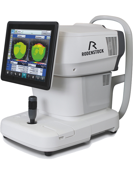
Tune-up your businessExperience 6 analyses in 1 device: Rodenstock Alino Multifunction Refractometer
Are you ready for something that is all-in-one, timesaving, space-saving and suits your business perfectly?Have a look at how powerful ALINO can be.6 in 1 solution01 RefractionHaving a good starting value for subjective refraction is essential. The QUICK REF MODE supports this even for uncooperative patients.02 KeratometryKAI (Kerato-asymmetry index) & KRI (Kerato-regularity index) give the first hint of possible irregularities on the cornea. This supports evaluation of the best vision correction or best-fitting contact lens.03 TonometryThe very gentle air puff created by a new generation of air-flow technology means that patients more readily accept the need for regular check-ups of the IOP measurement.04 PachymetryCorrecting the IOP by measuring the central corneal thickness is essential in interpreting the intraocular pressure.05 TopographyVarious topographic maps support you in carrying out vision screenings, contact lens fittings and in patient education.06 Dry EyeObserve the customers’ eye health and evaluate the long-term tolerability of contact lenses.Take advantage of the combination of tests:Additional servicesTonometry + PachymetryContact lens fitting andtolerance controlKeratometry + Topography + Dry EyeEye healthcareTopography + Tonometry + Pachymetry + Dry EyeSpecifications Rodenstock Alino Multifunction Refractometer
REFRACTIVE POWER MEASUREMENTSpherical refractive power (S) -30.00 D to + 25.00 D (at VD = 12.0 mm)Cylindrical power (C) 0.00 D to ± 12.50 D (at VD = 12.0 mm)Astigmatic axis (A) 0° to 180°KERATOMETRY MEASUREMENTCorneal curvature radius 5.00 mm to 13.00 mmCorneal astigmatic axis 0° to 180°INTRAOCULAR PRESSURE MEASUREMENTMeasurement range 1 mmHg to 60 mmHG (1 hPa to 80 hPa)PACHYMETRY MEASUREMENTMeasurement range 300 μm to 800 μmTOPOGRAPHY MEASUREMENTCorneal curvature radius 5.50 mm to 10.00 mmCorneal astigmatic axis 0° to 180°AUXILIARY FUNCTIONInterpupillary distance Measurement range: 20 mm to 85 mmCorneal diameter and pupil diameter Measurement range: 1.00 mm to 14.00 mmDry eye observation Blinking Analysis, Tear Meniscus height, Hyperemia, Meibomian glandsDATA MANAGEMENTInternal Database Integrated SD CardPrinter Integrated thermal printerData Output Type 3x USB 2.0, 1x LAN, 1x SD Card-slot, 1x WLANExport Format CSV, XML, JPG, PDFDIMENSIONS & ELECTRIC REQUIREMENTSDimensions WDH 312 × 491 × 450 mmWeight Approx. 23 kgVoltage 100 VAC to 240 VACFrequency 50/60 HzPower consumption 110 VA | Topcon KR-8800 Autorefractor Keratometer
The Topcon KR-8800 Autorefractor Keratometer features fast, easy and accurate measurements. The Topcon KR-8800 Autorefractor Keratometer also offers a 20% wider measurement range, a color LCD screen, and a 2.0mm minimum pupil diameter. Fast and accurate refraction and keratometry readings are also standard on the KR-8800, as well as a new color LCD screen. The Topcon KR-8800 Autorefractor Keratometer offers Rotary Prism measurement for enhanced accuracy. Other features offered on the Topcon KR-8800 Autorefractor Keratometer are simultaneous display of binocular readings, scenic fixation chart, USB connection for fast data transfer, and auto fire mode for fast reliable data collection.Simultaneous Display of Binocular Readings
The new color LCD monitor simultaneously displays the readings of both right and left eyes so that the operator can instantly observe all of the pertinent information.Wider Measurement Range
The new 8800 series’ wider range allows measurements to be obtained on patients with up to 10D of astigmatism.Rotary Prism Measurement System
The new 8800 series features Topcon’s exclusive Rotary Prism Technology for unmatched accuracy and reliability. The off-centered ring target is able to measure areas that may be occluded by a small diameter pupil. The prism angle enables a much wider area of the retina to be measured. As a result, the measurement image provides extremely reliable data.Faster Measurement Topcon KR-8800 Autorefractor Keratometer
The 8800 series are among the fastest auto refractors in the world. A specially designed CPU enables the 8800 series to significantly reduce the measurement time.Scenic Fixation Chart
The scenic color chart makes eye fixation easier and is adjustable in two brightness levels for optimal measuring results with either large or small pupils.Auto Measurement
As soon as the KR-8800 is properly aligned, this innovative function initiates the measurement process and completes three readings of each eye providing that proper alignment is maintained.Cornea Diameter Measurement
Corneal diameter can be measured either during testing of the patient or later by recalling the image of the cornea on the monitor. |




Reviews
There are no reviews yet.