| Description | With the intraoral scanner dwio the canadian company Dental Wings presented at the IDS a really revolutionary approach in the field of intraoral scanner. The digitally interested users will have noticed the similarity of the dwio with the Steinbichler intraoral scanner which was launched at the IDS 2 years before. In fact, the dwio intraoral scanner represents a further development. | InMode’s EVOKE is the first and only FDA-cleared, all-in-one hands-free facial remodeling device. This state-of-the-art thermal skin rejuvenating platform remodels facial tissue and delivers the ultimate in thermal facial procedures. Facial remodeling is one of the top aesthetic procedures patients seek as they want to achieve a youthful appearance without invasive surgery and related concerns. Evoke’s proprietary non-invasive technology is an industry-first in delivering a structural re-organization of the facial and neck tissues resulting in a three-dimensional remodeling. The treatment rejuvenates your face, neck, chin, cheeks and jowls with no anesthesia, surgical scars or downtime. | This system allows not only a quantitative nutational infrasonic infiltration, but also a nutational infrasonic liposculpture.Manufactured for a safe liposculpture respecting the non-fatty tissues. | Deka Synchro Replay Laser Nd-Yag Standard Handpieces – More than 10 selectable handpieces
– Different spotsizes available (2.5 – 24 mm)– Automatic spot recognition system– Ergonomic connector for air-cooling devices | Energist NeoGen PSR offers a range of treatment options, and delivers the unparalleled results your patients expect: The unique energy delivered by NeoGen is non fractionated and not dependant on a chromophore for its uptake. This provides uniform energy absorption, ensuring consistent treatment of the skin, and at high energies supporting significant tightening. | Treatments offered with the InMode OptimasMorpheus8- Skin Resurfacing and RejuvenationLumecca IPL – Age spot and sun spot treatmentForma – Skin remodeling at its bestAvailable Handpieces:
LUMECCA
FORMA
FRACTORA
DIOLAZEXL
VASCULAZE
VLAZE
MORPHEUS8LUMECCA
Fastest IPL Results*
*Clearance in 1-2 sessions, compared to 4-5 sessions with competing devices |
| Content | Dwio Intraoral Scanner Dental WingsIntraoral scanner
Based on a novel and incredibly compact 3D capture technique called Multiscan Imaging, the Dental Wings intraoral scanner allows dentists to take digital impressions in a natural, fluid manner. Its remarkably small handpiece, gesture control, and easy maintenance make it a compelling digital impression option for clinicians.Design approach: the patient first
The intraoral scanner was designed to address the needs of dental professionals looking for an intuitive technology for capturing digital impressions. With the overall objective to help dentists focus on the patient rather than the technology, the Dental Wings intraoral scanner offers:– A remarkably small handpiece very similar in size, shape, and weight to existing dental handpieces. Its familiar shape allows to assume a natural position relative to the patient.
– Visual and auditive feedback — a luminescent ring and audible signals indicate when data is being successfully captured, allowing the user to focus in the mouth rather than on the screen.
– Easy maintenance of the lightweight all-metal handpiece that has no moving parts, no complex illumination or cooling system.
– Motion control technology for a no-touch operation of the system while the user has gloves on.Scanning technology Dwio Intraoral Scanner Dental Wings
Five miniaturized 3D scanners in the handpiece tip directly view the teeth and soft tissue from multiple orientations simultaneously, capturing even the most difficult to see areas of preparations with minimal effort by the user.Open workflows
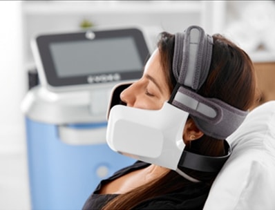
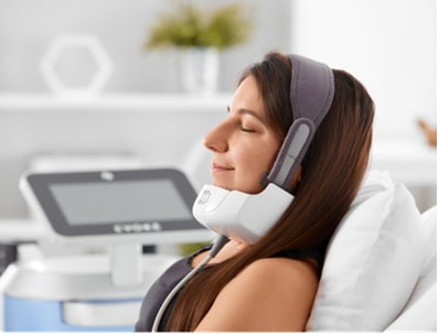
The intraoral scanner is a dedicated “scan only” chairside system with multiple options for the design and fabrication steps. Captured data is seamlessly transmitted to in-office or remote systems via DWOS Connect for prosthesis design and production. Users of Dental Wings’ simple to use yet powerful DWOS CAD suite of design applications, as well as other open dental CAD software, are able to import the scan data directly. | Inmode Evoke Face & Neck Aesthetic TreatmentsInMode EVOKE proprietary non-invasive technology is industry-first in delivering a structural re-organization and contouring of the facial and neck tissues. The EVOKE non-surgical treatment remodels and contours your This hands-free design and programmable technology by InMode is non-invasive, the only platform to have temperature monitoring and automatic, user-programmable temperature on/off control. Its hands-free design and programmable technology make it an ideal treatment at this time of social distancing. This advanced thermal skin rejuvenating platform restructures facial tissue and delivers the ultimate in thermal facial procedures with no anesthesia, surgical scars or downtime.EVOKE requires only minimal patient-physician face- to-face contact during the procedure, making it easy to comply with COVID-19 related directives. EVOKE: Non-invasive and Safe
By using proven bipolar radiofrequency energy, EVOKE sub-dermally remodels the facial tissues to provide a more defined neck and jaw line. Its hands-free facial applicators deliver uniform and volumetric heating to the skin and subdermal layer.Using this innovative platform, our surgeons provide a variety of facial remodeling procedures specifically tailored to your requirements. Safety Features Inmode Evoke Face & Neck Aesthetic Treatments - Built-in real-time audible indicators that sound when the optimal temperature is reached, providing feedback on treatment progress
- A Patient Call Button to allow you to be in contact with your surgeon at all times.
- Treatment screen that enables physicians to visually monitor the thermal effect of each applicator at any point during the procedure
- Color-blind RF technology which ensures that the device can be safely and effectively used on all skin types
You can stay tension-free and relaxed – sleep, read a book or watch TV, while our skilled surgeons perform this non-invasive procedure. You can even talk while the treatment is going on. Treatment
During the Evoke non-surgical treatment, you will feel only a mild warmth. Some redness may be seen which subsides in a few hours. There is absolutely no recovery period or downtime associated with EVOKE. Patients can return to normal activities immediately after these quick and painless treatments. Come in during your lunch time and go back to work. It’s simple as that. | Euromi Eva SP7 Lipomatic Liposuction Unit{tab System composition}- 15-inch touch screen that can be angled from 0 to 45 degrees
- Excellent visibility for the surgeon during the procedure
- Control panel with touch screen allowing the infiltrated and aspirated zones to be visualized
- Control of the volume infiltrated and aspirated millimeter per millimeter in the zone selected on the touch screen
- The n.i.l technique ensures an homogeneous infiltration with a constant pressure, whatever the fat density
- Infiltration and aspiration data recording on USB flash memory
- Compatible with the lipofilling material n.l.f System 1 & 2
- In compliance with the European standards – CE marking
{tab Advantages}- Preservation of non-fatty tissues
- Very easy to change zones thanks to the mapping of the different zones on the touch screen
- Shorter post-procedure recovery
- Less traumatising (fewer hematomas and major reduction of ecchymosis and oedemas)
- Less painful for the patient
- Infrasonic frequency (25 Hz) comfortable for the patient
{tab Accessories} Euromi Eva SP7 Lipomatic Liposuction Unit- Evamatic/evamatic
- Control panel with touchscreen
- Quantitative infiltration system
- High-speed aspirator
- Complete set of cannulas
- Accessories
The Eva sp 7 allows a quantitative nutational infrasonic infiltration and also a nutational infrasonic liposculpture.Manufactured for a safe liposculpture respecting the non-fatty tissues, the system is entirely digitalizedIt complies with European standards (CE marking) and is compatible with N.L.F. System, our kit for lipofilling.This product is a medical device intended for use by qualified healthcare professionals.If you are a healthcare professional and would like more information about this product, please complete the form opposite.If you are an individual, for more information, please refer to your healthcare professional.Special features of Eva sp 7:- The screen provides excellent visibility for the surgeon during the procedure: infiltrated and aspirated areas.
- In addition, changing zones is very easy thanks to the mapping of the different zones on the touch screen. This screen allows you to control the volume infiltrated ml / ml in the zone selected on it.
- Infiltration and aspiration data can be saved on a USB drive.
| Deka Synchro Replay Laser Nd-Yag Inside Moveo Innovation Moveo HR: Focus on Hair Removal
The multiple passes of Moveo HR over small defined areas cause gradual heating and destruction of unwanted hairs.
Moveo PL: Focus on Pigmented Lesions
The new MOVEO PL is indicated for skin age and sun spots progressively heating the pigmentedMoveo SR: Focus on Skin Rejuvenation
The multipass controlled action of MOVEO SR causes a progressive increase in heat at the dermal level, which stimulates a neocollagenic process.Moveo VL: Focus on Vascular Lesions
The specific action of Moveo VL acts directly and effectively on vascular redness.The versatile FT lamp can be added to the system at any time, enhancing the user’s ability to treat superficial Vascular and Pigmented Lesions.The FT pulsed light has ten different filters divided in five spectra of emission (500-520-550-600-650nm) and two spot areas. This flexibility is useful to adapt the emitted light to the patient skin type, application and treatment area.Deka Synchro Replay Laser Nd-Yag Standard Handpieces – More than 10 selectable handpieces
– Different spotsizes available (2.5 – 24 mm)– Automatic spot recognition system– Ergonomic connector for air-cooling devicesDeka Synchro Replay Laser Nd-Yag
Wavelength: 1064 nm
Pulse Energy: 120 J (max)
Pulse Lenght: 0.2 ms to 300 ms
Frequency Single Pulse: 10Hz (max)
Spot Diameter: 20mm, 15mm, 10mm, 7mm, 5mm, 2.5mm
Emission Spectrum: 500-950 nm and 650-950 nmTo Include:
1 x Foot Switch
1 x Key
1 x Module UPL Handpiece
6 x Deka Laser Handpieces
1 x ManualInside Moveo InnovationMoveo HR: Focus on Hair Removal
The multiple passes of Moveo HR over small defined areas cause gradual heating and destruction of unwanted hairs.
Moveo PL: Focus on Pigmented Lesions
The new MOVEO PL is indicated for skin age and sun spots progressively heating the pigmented lesions.Moveo SR: Focus on Skin Rejuvenation
The multipass controlled action of MOVEO SR causes a progressive increase in heat at the dermal level, which stimulates a neocollagenic process.Moveo VL: Focus on Vascular Lesions
The specific action of Moveo VL acts directly and effectively on vascular redness. | Energist NeoGen PSR LaserNeoGen PSR SystemNeoGen nitrogen plasma technology delivers controlled heating to the skin architecture stimulating a significant physiological response, without creating an open wound. Unlike ablative technologies there is no epidermal vaporisation or charring caused at time of treatment.
Unique TechnologyNeoGen converts nitrogen gas into plasma energy, the fourth state of matter. The plasma emerges from the handpiece in controlled pulses and rapid heating of tissue occurs as it gives up its unique thermal energy to tissue.
No Open WoundsThe treated photodamaged layers undergo a controlled thermal modification, while remaining intact, creating a natural dressing to provide protection and speed healing. at high energies the epidermis sheds, but only after a new, healthier skin architecture is formed beneath.
Treats the Whole Skin ArchitectureTreating the entire skin structure ensures optimal results as the entire surface is regenerated with associated neocollagenesis and neoelastogenesis. No islands of untreated skin remain. Clinical studies show significant tightening – Mean 22% improvement in upper eyelid tightening (JOCD 7, 169-179, 2008).
Eyelids and Periorbital TreatmentEnergist NeoGen PSR is the ideal option for the treatment of the upper and lower eyelids, as well as the wider periorbital region. Other technologies cannot be used in this area or deliver inconsistent results.
Long Lasting ResultsClinical studies show neocollagenesis and reduction in elastosis continuing for more than one year post treatment.The Proven Power of Plasma for Skin Regeneration Energist NeoGen PSR Laser• Unique technology – controlled pulses of nitrogen plasma energy
• Treats the whole skin architecture – no islands of untreated skin remain
• Deep Tissue Remodelling – epidermis acts as natural dressing post treatment
• Versatile – high or low energy treatment choices to fit your patients’ needs and lifestyle
• Precise – treat eyelids effectively with significant results
• Long-lasting effects – intense fibroblast activity supports continued regeneration.
• Consistent – non-fractionated and non-chromophore dependence assures uniform energy absorption
• Proven Plasma Skin Regeneration is supported by numerous IRB-controlled studies
TechnologyPlasma skin regeneration was introduced with a level of scientific and clinical testing never before seen in the industry:
• 3+ years of pre-clinical and clinical testing
• 16 separate studies
• more than 450 clinical study treatments –No scarring or Hypopigmentation noted in studies
• one year clinical histology
This level of testing is very rare in the industryEnergist NeoGen PSR uses a highly energized gaseous state known as plasma to produce a unique thermal profile to penetrate the superficial and deeper levels of the dermis to replace damaged collagen and encourage new collagen growth for new skin. Plasma is produced inside the handpiece through the combination of inert nitrogen gas and ultra-high frequency RF.The Energist NeoGen PSR is emitted in millisecond pulses; the longer the pulse, the more energy is delivered. Nitrogen molecules impact the skin’s surface and immediately transfer energy upon contact. When the nitrogen plasma impacts the skin surface, energy is immediately transferred to the tissue. Oxygen is purged from the skin surface by the plasma, preventing carbonisation. The energy delivered to tissue ranges from 1 – 4 Joules and is user-adjustable. The repetition rate of the plasma pulses can also be controlled, and ranges from 1 – 2.5 Hertz.Specifications Energist NeoGen PSR| Power: | 100 – 120 / 230 Vrms – 50/60 Hz – 650 Va | | Energy Output: | Pulsed nitrogen plasma – 1-4 Joules. | | Repetition Rate: | 1.0 – 2.5Hz | | Dimensions: | 470 x 430 x 1060mm | | Gas Requirement: | Medical Grade Nitrogen | | System Weight: | 18kg/40lb | | Trolley Weight: | 25kg/55lb (excludes gas tank) | | Ambient Environment: | 0 to 40 celsius | | Relative Humidity: | 30 to 75% | | atmospheric pressure: | 700 to 1600 hpa |
| Inmode Aesthetics Optimas with Lumecca 515 & 580LUMECCA
SR515
Spot size:10x30mm
Pulse Repetition:1pps at all energies
Wavelength:515-1200nm
Fluence:10-30J/cm2
Peak Power:High Peak Power
Pulse Duration:Short/Long
Cooling:Controlled
SR580
Technology:IPL
Spot size:10x30mm
Pulse Repetition:1pps at all energies
Wavelength:580-1200nm
Fluence:10-30J/cm2
Peak Power:High Peak Power
Pulse Duration:Short/Long
Cooling:Controlled
FORMA
RF Output Frequency: Up to 65 W
Output Frequency: 1 MHz
Temperature Cut-Off: Real time temperature measurements set by operator up to 43°C
FRACTORA
24 Pin:Type – Full Thickness Dermal; Configuration – 6×4; Pin Length – 3000u; Ablation Depth – 3 mm; Heating Depth – Between 3-5 mm; Max Output Energy 62 mj/pin; Output Frequency – 1 MHz; Repetition Rate – Up to 2 pps
24 Pin Coated:Type – Deep and Subdermal; Configuration – 6 x 4 Coated; Pin Length – 3000 u, Ablation Depth – 3 plus mm; Heating Depth – Between 3-5 mm; Max Output Energy 62 mj/pin; Output Frequency – 1 MHz; Repetition Rate – Up to 2 pps
60 Pin:Type – Mid Dermal; Configuration – 10×6; Pin Length – 600u; Ablation Depth – 0.6-1 mm; Heating Depth – Between 3-5mm; Max Output Energy 62 mj/pin; Output Frequency – 1 MHz; Repetition Rate – Up to 2 pps
DIOLAZEXL
Laser Wavelength:810 nm (diode)
Spot Size:12mm x 26 mm
Fluence:Up to 40 J/cm2
Pulse Duration:Short/Long
Light Cooling Guide:2-15°C
Repetition Rate:Single and Auto-Repeat
VASCULAZE
Laser Wavelength:1064 nm (diode)
Spot Size:3mm x 4mm
Fluence:40-300 J/cm2
Pulse Duration:5-100 msec
Light Cooling Guide:7-12° C
VLAZE
Laser Wavelength:1064 nm (yag)
Spot Size: 4 mm x 5 mm
Fluence: Up to 300 J/cm2
Pulse Duration: 5 ms 25 ms
MORPHEUS8
Type:Deep and Subdermal
Pin Length:4000
Ablation Depth:5mm +
Max Energy:62 mJ/pin at 75 W
Frequency:1 MHz
Repetition Rate:Up to 2 pps
A FULL BEAUTY SUITE
Optimas will help you optimize results for your patients and your businessProfessional grade aesthetic procedures – Optimas is a great suite of innovations in one workstation to help you achieve your goals. The Optimas full beauty suite has state-of-the-art light, laser, and radiofrequency devices for skin care and hair removal.Available Handpieces : Inmode Aesthetics Optimas with Lumecca 515 & 580
LUMECCA
FORMA
FRACTORA
DIOLAZEXL
VASCULAZE
VLAZE
MORPHEUS8LUMECCA
Fastest IPL Results*
*Clearance in 1-2 sessions, compared to 4-5 sessions with competing devicesLumecca is a breakthrough intense pulsed light (IPL) that delivers up to 3X more energy in the 500-600 nm range to improve efficacy for vascular and pigmented lesions. It is optimized for clinicians to treat a variety of skin types and conditions with just a single session.SR515 – Removal of Pigmented Lesions on Light Skin, Removal of Superficial Vascular Lesions
SR580 – Removal of Pigmented Lesions on DARK SkiN, Removal of DEEP Vascular Lesions, Removal of Vascular Lesions on DARK SkinFORMA
Face Remodeling.
Stimulates the formation of new collagen and improves the skin’s elasticity for long-lasting and remarkable resultsForma is the first auto-adjusting, non-invasive, thermal skin treatment for deep and uniform tissue stimulation. Forma uses radio-frequency power that flows uniformly between the electrodes to provide a comfortable thermal experience with immediate and subsequent contraction. There is no downtime in this effective lunch-time procedure that will make anyone look years younger!Acquire: Forma has a temperature sensor built into the hand piece which reads skin surface temperature 1000 times per second, allowing clinicians to acquire skin temperature in real time.
Control: Software algorithm allows unprecedented safety of RF delivery. The cut off temperature” feature reduces RF energy automatically when the hand piece senses that the required skin temperature has been reached.
Extend: Clinical evidence suggests prolonged exposure to temperature above 40°C is advantageous for optimal clinical outcomes. Only InMode’s A.C.E. technology allows you to utilize therapeutic temperatures safely and efficiently.FRACTORA Inmode Aesthetics Optimas with Lumecca 515 & 580
Radio Frequency MicroNeedling.
Fractora is a fractional skin resurfacing and subdermal tissue coagulation device that bridges the gap between fractional lasers and surgical procedures. Clinical papers demonstrate outcomes such as: improvement in skin complexion, reduction in skin irregularities and restoring skin to a more youthful appearance. This all occurs within one session or multiple sessions, depending on patient preference. Fractora can be used on active cystic acne and acne scars.Addresses multiple problems, including treatment of wrinkles with fractional coagulation and ablation.
Significant resolution of cystic acne and scars (improving acne scarring by 50%).
Safe on skin type VI with little risk of post inflammatory hyperpigmentation (PIH) which is common with other resurfacing methods.
Combination of fractional coagulation and volumetric heating through tips with various depths and pin density configurations.High efficiency of vascular and pigmented lesions due to high peak power and optimized output.
Complete photo rejuvenation in 1 or 2 treatments versus 4-6 treatments with standard IPLs.
Reduces treatment time thanks to large spot size and high pulse repetition rate.
Strong sapphire cooling tip results in painless procedure.
Fast pulse repetition rate at all settings. Highest peak power 3300W*DIOLAZEXL
Diode Hair Removal.
Gold Standard Diode Hair Removal Without Compromising Peak Power, Speed and Large Spot SizeDiolazeXL is the most effective at combining high peak power and a large spot size simultaneously in the same session. Clinicians can benefit from this first and only combination of speed, efficacy, safety and comfort to optimize their hair removal revenue.Gold standard wavelength, pulsing and power for optimal results and maximal safety.
Reduction in clinician treatment time – Powerful enough to target and treat even the most stubborn hair.
Virtually painless due to strong built in cooling.
Can treat up to skin type VI.VASCULAZE
Vascular Lesion Removal.
Get the ultimate in vascular resolution with a fast and chilled leg and facial vein treatmentVascular lesions (including angiomas, telangiectasias, port wine stains and leg veins) can be treated with the Vasculaze. The Vasculaze is optimized with high peak power that targets appropriate hemoglobin, so patients can benefit from a fast and safe treatment.Narrow and ergonomic spot size is optimized for vascular lesion treatment. High peak power. Strong contact cooling. No disposables and consumables.VLAZE
Vascular Lesion Removal.
Fast, powerful and ergonomic laser device. Treats vascular lesions such as veins, spider veins and port wine stains safely, comfortably and effectively on all skin types from very light to darker skin tones.MORPHEUS8
THE FIRST FULL BODY FRACTIONATED TECHNOLOGY
Fractional Remodeling and Contouring At Its BestThe Morpheus8 is a new subdermal adipose remodeling device (SARD) that fractionally remodels and contours the face and body. Penetrating deep into the skin and fat, this morphs the aging face of body into a more desired smooth and sleek appearance, for all skin tones.Deep and safe fractional treatment penetrating 4000 microns with an additional thermal profile of 1000+ microns
Extremely uniform effect
Little to no thermal damage to dermis
Safe on skin type VI with little risk of post inflammatory hyperpigmentation (PIH) which is common with other resurfacing methods. |


Reviews
There are no reviews yet.