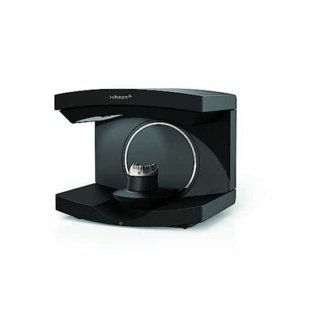
New 3Shape E4 – Ultimate productivity with our fastest scanner ever 3Shape is renowned for making very fast scanners, and the E4 is the fastest and most accurate scanner we have made to date. 4 x 5 MP cameras enable very high accuracy, powerful impression scanning and scanning of dies while they reside in the model to reduce scan steps and save time.
For those seeking the speed and accuracy to drive lab productivity and growth, look no further. The E4 boasts double the speed, double the accuracy, and double as many cameras as its predecessor the E3, delivering improved efficiency and consistency with every scan. From small labs seeking the best-of-the-best, to high-volume, full-service labs striving for a leaner workflow, our E4 lab scanner is the right choice for you.
Double the speed
The E4 scans a full arch in 11 seconds which is 2 times faster than the E3’s 22 seconds.
Double the accuracy
The E4 delivers accuracy of 4 microns compared to the E3’s 7 microns accuracy.
Double the cameras
4 x 5 MP cameras enable scanning of dies in the model omitting extra steps for scanning dies individually.
High precision impression scanning
Enabled by the scanner’s 4 x 5 MP cameras.
All the features from E1, E2, E3
Includes Auto-start scanning and articulator holder support.
3Shape E4 Premium Orthodontic Scanner Specifications:
Camera : 4 x 5 MP
Accuracy (ISO 12836) : 4 μm
Scan speed (full arch) : 11 sec
Scan speed (full arch impression) : 45 sec
Texture : Color
Scanning strategy : Die-in-model
Designed with a signature red ring, Generation Red E scanners provide optimized productivity, at no extra cost.
Intelligent Machine. Effortless Production. Superior Results.
The DGSHAPE DWX-42W enables wet milling of popular glass ceramic and composite resins with high precision and accuracy.
High speed production with proprietary DGSHAPE spindle
Expanded 6-tool Automatic Tool Changer for uninterrupted milling
LAN capable for easy connectivity
Included VPanel for easy monitoring of milling jobs and tools
iTero HD2.9 simply allows to digitally scan a prepared tooth and wirelessly send this information directly to the dental laboratory for fabrication of a CAD/CAM crown without the gooey mess.
SuniRay 2 achieves maximal image quality with superior diagnostic capabilities while maintaining the lowest radiation levels among all digital sensors. The comprehensive and feature-rich Prof. Suni Advanced Imaging Software supplements the SuniRay 2’s high-quality imaging with a wide array of user-friendly customization and image-enhancement tools. With the SuniRay 2, Suni has created a complete digital radiography system that seamlessly integrates with your entire practice.
From general to littles, this machine has you covered. This digital system excels at both general and pediatric practices, with great strengths in cosmetic dentistry and orthodontics as well.
Image Quality
According to DEXIS, their digital xray system provides detailed image quality and includes image enhancement tools that increase diagnostic capabilities. When the 9 evaluators were asked to rate the system’s image quality and enhancement aids, 89% rated them as excellent to very good. One of the evaluators commented that the system’s “ClearVu and image magnification allow us to diagnose very effectively.”Overall Satisfaction
When asked to rate their overall satisfaction with the DEXIS Digital X-ray System, 89% of the evaluators rated it as excellent to very good. Commented one evaluator, “DEXIS has been very responsive to our needs.”
Content
3Shape E4 Premium Orthodontic Scanner
New 3Shape E4 – Ultimate productivity with our fastest scanner ever 3Shape is renowned for making very fast scanners, and the E4 is the fastest and most accurate scanner we have made to date. 4 x 5 MP cameras enable very high accuracy, powerful impression scanning and scanning of dies while they reside in the model to reduce scan steps and save time.For those seeking the speed and accuracy to drive lab productivity and growth, look no further. The E4 boasts double the speed, double the accuracy, and double as many cameras as its predecessor the E3, delivering improved efficiency and consistency with every scan. From small labs seeking the best-of-the-best, to high-volume, full-service labs striving for a leaner workflow, our E4 lab scanner is the right choice for you.Double the speed
The E4 scans a full arch in 11 seconds which is 2 times faster than the E3’s 22 seconds.Double the accuracy
The E4 delivers accuracy of 4 microns compared to the E3’s 7 microns accuracy.Double the cameras
4 x 5 MP cameras enable scanning of dies in the model omitting extra steps for scanning dies individually.High precision impression scanning
Enabled by the scanner’s 4 x 5 MP cameras.All the features from E1, E2, E3
Includes Auto-start scanning and articulator holder support.3Shape E4 Premium Orthodontic Scanner Specifications:
Camera : 4 x 5 MP
Accuracy (ISO 12836) : 4 μm
Scan speed (full arch) : 11 sec
Scan speed (full arch impression) : 45 sec
Texture : Color
Scanning strategy : Die-in-model3Shape E4 Premium Orthodontic Scanner Package includes:
– Object Scanner
– (3) Interface Plate
– Scan Height Adaptor
– Articulator Calibration
– Object Articulator
– Transfer Plate of choice
– 2-Cast Fixture
– Wax-up Fixture
– Software Package: ScanIt Orthodontics, 3Shape Communicate Inbox, Ortho Analyzer, Study Model Builder (Incl. STL output)
3Shape E4 Premium Orthodontic Scanner Uses and Advantages
20% faster scanning than the previous generation
Next generation platform: E1, E2 and E3 upgraded to the same next generation platform as E4.
High precision impression scanning, articulator scanning and auto start.
The DWX-42W enables wet milling of popular glass-ceramic composite resins with high precision and accuracy. High-Speed Production with proprietary DGSHAPE spindle. Expanded 6-tool Automatic Tool Changer for uninterrupted milling. LAN capable for network and hardware connection and communication. VPanel user interface for easy monitoring of milling jobs and tools.The DGSHAPE DWX-42W enables wet milling of popular glass ceramic and composite resins with high precision and accuracy.
High speed production with proprietary DGSHAPE spindle
Expanded 6-tool Automatic Tool Changer for uninterrupted milling
LAN capable for easy connectivity
Included VPanel for easy monitoring of milling jobs and tools
iTero HD2.9 simply allows to digitally scan a prepared tooth and wirelessly send this information directly to the dental laboratory for fabrication of a CAD/CAM crown – without the gooey mess. The wireless transmission saves shipping time and eliminates distortion that can often be found with traditional stone modelsThe digital scan is, quite literally, digitally perfect. We then deliver the digital scan to our lab where a precision fitting restoration is created.iTero HD2.9 system eliminated the uncomfortable patient experience of traditional impressions, utilizes advanced state of the art digital technology, and provides with superior fitting restorations with little or no adjustmentsiTero Intra-Oral Scanner
Model: HD2.9
2014 Date of Mfg.
Brochure/Video: Click Here
Limited Use
Includes:
iTero Software (pre-loaded)
90 Day Parts Warranty
User Manual(s)
Free Standard Shipping (Commercial USA Addresses)
Its ergonomic design comes with safe docking station. It has thin fiber optic cable which enables quick data transfer from the wand to the cart. This optimized cable length assists various cart positions in the operatory.Orthodontic treatment is more than just straightening teeth; it’s about ensuring your teeth and jaws are in alignment. To achieve this, we need to get an accurate look at where your teeth are positioned. To do this, we use the iTero Intraoral digital scanner.A good impressionIn the orthodontic and dental industries, ‘impressions’ is the term we use for getting a good picture of where your teeth currently sit. These impressions were previously taken using a type of dental cement. Pasted on your teeth, patients were required to lie for a few minutes while the cement-like material set. The dental professional could then remove the set material which had formed an ‘impression’ of your teeth.This process was messy, rather unpleasant and not very comfortable. Fortunately, that method is a thing of the past. These days, we use scanning technology and at L&L Orthodontics, our technology of choice is the iTero Intraoral digital scanner.
What is the iTero scanner?
The iTero scanner is new 3D orthodontic scanning technology. Using the latest developments in intraoral scanning, the iTero scanner accurately scans your teeth as part of your orthodontic assessment. The iTero scanner is made up of a small, wand-like device which is used to scan your teeth and jaws. The data it is collecting then appears on the computer screen, allowing you to see the ‘impressions’ the scanner is taking.The iTero scanner works in with Invisalign®* treatment and lets you see what your teeth will look like post-treatment, even before you’ve started. Before we recommend a course of treatment for you, we’ll take impressions of your teeth using the iTero scanner to ensure we are making our recommendation on accurate information.To find out more about the iTero scanner and other orthodontic technology we use here at L&L Orthodontics, book your specialist consultation today.
SuniRay 2 Digital Intraoral Sensor Size 1 and Size 2
X-Ray Imaging Properties
Resolution: The pixel resolution of the high resolution (HiRes) sensors exceeds 15 lp/mm. The standard resolution of an X-Ray image, as measured by the modulation transfer function (MTF) exceeds 12 lp/mm. This is measured by using a standard 60 kV intraoral X-ray source.
Does Efficiency: The sensor will produce a high quality image with an X-Ray dose that is only a fraction of the standard dose required by Dental X-Ray film.
Diagnostic Efficacy: The system will produce a superior image quality of images obtained when using standard dental X-Ray film. This allows the dentist to diagnose standard intraoral pathologies encountered during screening procedures.
Wide dynamic range: The sensor pixel well capacity is very high, allowing a higher accommodation of greyscale (bone density) dynamic range.
Sensor Material Biocompatibility SpecificationsThe sensor’s body material and cable material are biocompatible. The sensors and all component materials have been tested to comply with ISO standards, and specifically, the sensor complies with EN IS0-10993 (Biological Evaluation of Medical Devices).
Technical data
Size 1
Size 2
Sensor Dimensions (mm)
39.5 x 26
43.5 x 31.5
Active Area (mm)
31.1 x 20.2
35.2 x 26.2
Sensor Technology
CMOS APS Fiber Plate
CMOS APS Fiber Plate
Maximum Gray Levels
4096
4096
Sensor Cable
3 feet (1m)
3 feet (1m)
Cable Attachment
Reinforced Strain Relief
Reinforced Strain Relief
USB Module
Integrated USB 2.0 Module
Integrated USB 2.0 Module
Software
Prof. Suni
Prof. Suni
X-Ray Imaging Properties SuniRay 2 Digital Intraoral Sensor Size 1 and Size 2
Resolution: The pixel resolution of the high resolution (HiRes) sensors exceeds 15 lp/mm. The standard resolution of an X-Ray image, as measured by the modulation transfer function (MTF) exceeds 12 lp/mm. This is measured by using a standard 60 kV intraoral X-ray source.
Does Efficiency: The sensor will produce a high quality image with an X-Ray dose that is only a fraction of the standard dose required by Dental X-Ray film.
Diagnostic Efficacy: The system will produce a superior image quality of images obtained when using standard dental X-Ray film. This allows the dentist to diagnose standard intraoral pathologies encountered during screening procedures.
Wide dynamic range: The sensor pixel well capacity is very high, allowing a higher accommodation of greyscale (bone density) dynamic range.
Soredex Cranex-D Panoramic X Ray
The Soredex Cranex D with cephalometric option provides a full range of digital panoramic and cephalometric dental x-ray examinations to meet the diagnostic needs of almost any general dental or specialty practice. With it’s minimal footprint, this Soredex option will help elevate the diagnostic functionality of any dental provider.Soredex Cranex D provides a full range of high quality digital panoramic x-ray examinations to meet the diagnostic needs of almost any dental practice – all at an affordable price.Acquisition computer included
Features Soredex Cranex-D Panoramic X Ray
CRANEX D is a direct digital panoramic and cephalometric x-ray system that provides superior image quality and more diagnostic value. Available as a single or dual sensor system, the CRANEX D grows with your practice, making it a valuable addition to meet the needs of your office. It combines quality images, ease-of-use, and flexibility to make your day-to-day diagnostics easy and accurate.Benefits
The system features superb anterior image quality in panoramic imaging with its patented Variable Panoramic Collimator (VPC) by expanding the focal trough by 50% compared to traditional techniques. Additional innovative features on the unit include Automatic Collimator Selector (ACS) to easily set proper collimation for the imaging program prescribed, as well as the Automatic Exposure Setting (AES) where the system recommends proper cephalometric exposure values based on the size of the patient. These automated and optimized exposure settings maximize the practice’s workflow. CRANEX D is built for contemporary, fast-paced dental care where reliability is key.The Soredex Cranex D with cephalometric option provides a full range of panoramic and cephalometric examinations to meet the needs of almost any practice.SOREDEX has over 30 years of expertise in state-of-the-art dental imaging systems. The Soredex® Cranex D with cephalometric option provides a full range of panoramic and cephalometric examinations to meet the needs of almost any practice.
Dexis Intraoral Digital Dental X-Ray SensorSoftware Ease of Use
According to the manufacturer, the DEXIS x-ray software was designed to be highly effective throughout diagnosis, treatment planning, and treatment. It is intended to serve as an imaging hub, managing all digital images, including intra- and extraoral radiographs and photographs. The manufacturer states that the images can be quickly organized, stored, retrieved, printed, and shared with patients and colleagues. The 9 dentists who participated in this product evaluation were asked to rate the DEXIS Digital Imaging software on its ease of use with regard to taking and accessing radiographs and for diagnosis, patient education, and communication with colleagues and insurance companies. Six of the evaluators rated the system as excellent and 3 rated it as very good. One evaluator commented that “when communicating with patients, we use DEXIS images displayed on a monitor in front of the patient every day on virtually every patient.”Software Integration
The manufacturer claims that the DEXIS imaging software can be easily integrated with all major practice management systems. According to DEXIS, the DEXIS Integrator automatically synchronizes with the patient record.When the evaluators were asked to rate the software integration with practice management software and interface with hardware (eg, cameras, panoramics, and scanners), 78% rated it as excellent to very good, while 22% rated it as good. One of the evaluators said, “The bridge to our practice management systems is seamless. We use [the] DEXIS digital pan[oramic] so that we can store panoramic x-rays easily in the same location as full series. We use an intraoral camera to store preoperative and interoperative photographs for excellent documentation.”Sensor Comfort and Ease of Use
According to DEXIS, the heart of their digital system is the PerfectSize sensor. The manufacturer states that their single-size sensor has 3 advantages:
1) because the electronics are on the back of the sensor, the active imaging area is most of the total sensor size;
2) the beveled corners, slim profile, and rounded casing allow for accurate placement and patient comfort;
3) patented cord placement allows the practitioner to take vertical and horizontal bitewings and all periapicals with only 1 universally sized sensor.
The evaluators were asked to rate both the ease of use of the sensor and patient comfort. Five evaluators rated ease of use excellent and 4 rated it very good. With regard to patient comfort, 3 evaluators rated the sensor excellent and 6 rated it very good. One evaluator noted that he experienced a few issues when placing the sensor in some children and patients with very small mouths. Another evaluator, however, is very happy with the sensor, saying, “I chose DEXIS to take a full radiographic series on every patient. It was the only direct image system we tested that met that objective.”Image Quality
According to DEXIS, their digital xray system provides detailed image quality and includes image enhancement tools that increase diagnostic capabilities. When the 9 evaluators were asked to rate the system’s image quality and enhancement aids, 89% rated them as excellent to very good. One of the evaluators commented that the system’s “ClearVu and image magnification allow us to diagnose very effectively.”Overall Satisfaction
When asked to rate their overall satisfaction with the DEXIS Digital X-ray System, 89% of the evaluators rated it as excellent to very good. Commented one evaluator, “DEXIS has been very responsive to our needs.”System Includes* a highend CCD sensor* portable PC card and DEXusb connection options* DEXIS imaging software* a complete set of sensor holders and accessories* on-site training* a printed instruction manual* training tutorials on CD* a quick reference guide for the operatory* 1 year of DEXcare support.


Reviews
There are no reviews yet.