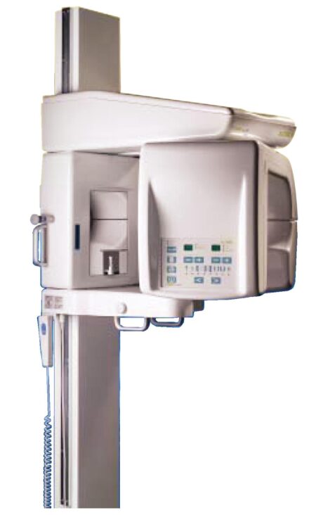
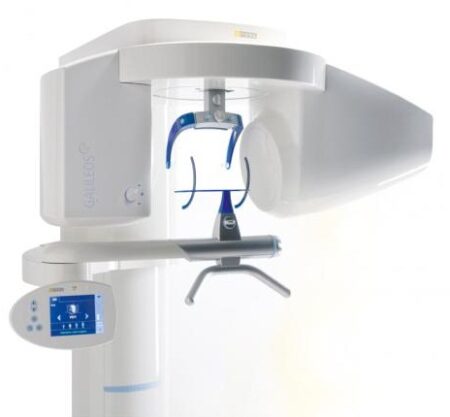
| Description | With the integrated FaceScanner in GALILEOS you can now record the facial surface parallel to the x-ray image. And that, as opposed to traditional face scanners without the use of lasers, which is therefore gentler on the patients. Thanks to the simultaneous x-ray image and surface depiction, the fully automated overlaying of the data is extremely precise. In addition, texture and coloring in the software allow for a particularly realistic depiction of the soft tissue proportions – an ideal tool for patient consultation! | Sirona Galileos Dental Cone Beam Imaging System The system captures images using the lowest possible radiation dose and is designed to provide high-quality images for implant planning, treatment guide design, jaw function analysis and more. | Caries diagnostic aid for surface caries.
TRIOS 4 has built-in fluorescent technology that aids in the identification of possible caries. Using TRIOS 4, dental professionals can now be aided in the early-detection of surface caries, without the need for an additional scanning device.
Caries diagnostic aid for interproximal caries.
TRIOS 4 will also feature a dedicated transillumination smart tip later this year. The smart tip will aid in the identification of possible interproximal caries undetectable to the eye, without emitting radiation. | SuniRay 2 achieves maximal image quality with superior diagnostic capabilities while maintaining the lowest radiation levels among all digital sensors. The comprehensive and feature-rich Prof. Suni Advanced Imaging Software supplements the SuniRay 2’s high-quality imaging with a wide array of user-friendly customization and image-enhancement tools. With the SuniRay 2, Suni has created a complete digital radiography system that seamlessly integrates with your entire practice. | TRIOS Color automatically measures the teeth shades as you scan. The dentist has the option to evaluate and highlight relevant areas for the lab. The complete shade information sis sent to the lab together with the impression. | WaterLase iPlus includes the all-new SureFire™ delivery system, with redesigned optics that can more efficiently deliver precise amounts of Er,Cr:YSGG laser energy to cut enamel, dentin and bone. The SureFire delivery system is optimized to provide the dentist with more up time with a replaceable, disposable shield that protects the vital optical components in the fiber, resulting in greater dependability. |
| Content | Sirona GALILEOS Comfort/Compact| GALILEOS Comfort | GALILEOS Compact | | Oral and maxillofacial surgeons
Dental clinics
Private clinics
Orthodontists | General practioners
Dental implant practices
Oral surgeons |  | | | Comparison Table of Galileos Comfort/ Compact & Orthophos XG 3D X-Ray systems | | Technical overview | GALILEOS Comfort | GALILEOS Compact | ORTHOPHOS
XG 3D | | Field of view | (15 x 15 x 15) cm³ | (12 x 15 x 15) cm³ | 8 cm x 8 cm (Ø x Höhe) | | 3D Resolution (isotropic voxel size) | 0.3/0.15 mm | 0.3 mm | 0,2 mm; 0,1 mm (available at the end of 201) | | Scan time / exposure time | 14 s / 2 – 6 s | 14 s / 2 – 6 s | 2–5 s | | Reconstruction time | 2.5 – 4.5 min | 4.5 min | 4,5 min | | Patient position | standing / seated | standing / seated | Standing/seated, chin rest/bite block, occlusal bite block for automatic patient positioning with 2D PAN images | | X-ray generator | | kV | 85 | 85 | 60–90 | | Effective dosage | 29 µSv / 68 µSv **
(21 mAs, 85 kV) | < 29 µSv / 68 µSv **
(21 mAs, 85 kV) | 43–175 μSv (Schulze)
(Standard: 100 μSv) | | Space requirements (minimum) | 1,6 x 1,6 x 2,25 m (d x w x h) | 1,6 x 1,6 x 2,25 m(d x w x h) | 1,5 m x 1,1 m x 2,25 m (Pan ), | | Space requirements (recommended) | >1,8 x 1,8 x 2,5 m (d x w x h) | 1,8 x 1,8 x 2,5 m (d x w x h) | 1,7 m x 1,3 m x 2,5 m (Pan ) | | User Interface | Full color touchscreen | Multipad | EasyPad | | Patient fixation | Occlusal bite block, forehead | Occlusal bite bock, forehead | | | Positioner | support, chin rest, head positioner *** | support, chin rest | | | Upgradable to Comfort version | | | | | Optional floor stand | | | | | Wheelchair accessible | | | | | Remote control | | | | | Programs | | 150 µm resolution for close-up reconstruction | | | | | 300 µm resolution for standard volumes | | | | | High contrast mode for optimized hard tissue display | | | | | Views | PAN with 3D slice navigation | PAN with 3D slice navigation | Tiltable 2D slices, custom | | TSA, axial, sagittal, coronal, CEPH lat., CEPH pa/ap | TSA, axial, sagittal, coronal | 3D slicing, TSA, LSA, axial, sagittal, coronal | | 3D model, 1-click OP reports | 3D model, 1-click OP reports | 3D model, implant-oriented view, 1-Click OP reports |
With the integrated FaceScanner in GALILEOS you can now record the facial surface parallel to the x-ray image. And that, as opposed to traditional face scanners without the use of lasers, which is therefore gentler on the patients. Thanks to the simultaneous x-ray image and surface depiction, the fully automated overlaying of the data is extremely precise. In addition, texture and coloring in the software allow for a particularly realistic depiction of the soft tissue proportions – an ideal tool for patient consultation! |
| SIRONA GALILEOS COMFORT PLUS
GALILEOS Comfort Plus is an advanced CBCT that provides seamless work flow integration. With an optional HD mode and a 14 second scan, the GALIELOS offers clinicians and specialists numerous options for diagnosis, treatment and patient consultation with superior image quailtiy and low radiation dose. It includes Integrated Implantology and FaceScan, which helps patients better understand and accept treatment recommendations. SICAT Function enables diagnosis and treatment of TMD, and SICAT Air (coming soon) software allows you to anyalze airways.Features:High-Quality Images:
This dental imaging system is able to position itsef and capture a superior quality of images with each scan, ultimately ensuring that you are receiving the detailed images you need, every time.Panoramic:
This imaging system provides crisp panoramic images to your practice with sensors built for high-quality 2D images. With multiple modes, it’s meant to give you the images you need.3D Conebeam:
Equipped with a high-quality, 3D sensor, this machine is capable of producing crisp and detailed representations of what you need to see. It’s cross-sectional images provide you with the high precision you need.Medium Field of View:
The 15×12 cm field of view option on this machine function allows for the versatility of a standard field of view.Sirona Galileos Dental Cone Beam Imaging System /Full Complete System Software and Hardware :
– Phantom
– Workstation
– Display Monitor
– Software
– ManualsAccessories
– 3D
– Galileos Comfort includes the following 3D views: pan with 3D slice navigation, TSA, axial, sagittal, coronal, 3D model, implant alignment and one-click op reports.
– MEDIUM FOV
– The system’s ellipsoid field of of view (15 x 12 cm) captures the complete dentition, as well as the ascending rami and sinus regions.
– ADJUSTABLE FOV
– Adjustable collimation can save time and reduce patient exposure by allowing you to scan only the regions of interest.
– STANDING DESIGN
– The unit features standing, face-to-face image programming as well as a patented head positioner, ensuring patient comfort and proper positioning.Sirona Galileos Comfort PlusCompact Model D3437Approximately 1100 CT images takenOperating PC includedFOV of the Gallileos Compact is 12×12 cmCrystal Clear 3D/CT imagesSeamless Cerec Integration | 3Shape TRIOS 4 Intraoral ScannerOur most powerful intraoral scanner to date Take your dentistry beyond treatment
Our intraoral scanning solutions allow you to take your dentistry beyond. The new 3Shape TRIOS 4 combines our established superior scanning technology with groundbreaking caries diagnostic aid technology* for both surface and interproximal caries. No additional scanning device needed. Smart tips 3Shape TRIOS 4 Intraoral ScannerThe new generation of TRIOS smart tips feature instant-heat technology for optimized scanning.Be scan-ready in seconds with instant-heat technology.Scan 2-3 times as many patients with instant-heat in combination with 30% more battery life.Optimal scanning due to automatic use counter and tip-change alert.The TRIOS system is a complete in-house ecosystem including a scanner and extensive design software. This means there are plenty of options depending on your needs. Those who are just looking for a scanner and want to utilize labs may look into the basic, cheaper options. Those who want to design crowns, bridges and other prosthetics can opt for design modules. The only missing component is a milling machine, which if you want to mill restorations in-house, will have to be from a third party. This proves to be a weak link in the digital chain.At iDD, we have been using the TRIOS 3 for over four years in our clinic, spending literally thousands of hours learning the workflow. We have carried out all aspects of dentistry using the TRIOS system including same-day crowns, implant surgical guides, implant restorations and digital smile design.Disclaimer: This is an impartial review of the scanner and we do not have any ties to the company. The team at iDD remain unwaveringly committed to providing you with objective and trustworthy information and therefore do not seek any sponsorship. 3Shape kindly loaned us a TRIOS 4 scanner, due to a personal interest in upgrading our older TRIOS 3 model. Our team at iDD are constantly approached by scanner companies to review their scanners and it is always made clear that they will have no part in writing reviews or restricting any conclusions iDD makes in our thorough analysis and clinical use of these products. | SuniRay 2 Digital Intraoral Sensor Size 1 and Size 2
X-Ray Imaging Properties- Resolution: The pixel resolution of the high resolution (HiRes) sensors exceeds 15 lp/mm. The standard resolution of an X-Ray image, as measured by the modulation transfer function (MTF) exceeds 12 lp/mm. This is measured by using a standard 60 kV intraoral X-ray source.
- Does Efficiency: The sensor will produce a high quality image with an X-Ray dose that is only a fraction of the standard dose required by Dental X-Ray film.
- Diagnostic Efficacy: The system will produce a superior image quality of images obtained when using standard dental X-Ray film. This allows the dentist to diagnose standard intraoral pathologies encountered during screening procedures.
- Wide dynamic range: The sensor pixel well capacity is very high, allowing a higher accommodation of greyscale (bone density) dynamic range.
Sensor Material Biocompatibility SpecificationsThe sensor’s body material and cable material are biocompatible. The sensors and all component materials have been tested to comply with ISO standards, and specifically, the sensor complies with EN IS0-10993 (Biological Evaluation of Medical Devices). | Technical data | Size 1 | Size 2 |
|---|
| Sensor Dimensions (mm) | 39.5 x 26 | 43.5 x 31.5 |
|---|
| Active Area (mm) | 31.1 x 20.2 | 35.2 x 26.2 |
|---|
| Sensor Technology | CMOS APS Fiber Plate | CMOS APS Fiber Plate |
|---|
| Maximum Gray Levels | 4096 | 4096 |
|---|
| Sensor Cable | 3 feet (1m) | 3 feet (1m) |
|---|
| Cable Attachment | Reinforced Strain Relief | Reinforced Strain Relief |
|---|
| USB Module | Integrated USB 2.0 Module | Integrated USB 2.0 Module |
|---|
| Software | Prof. Suni | Prof. Suni |
|---|
X-Ray Imaging Properties SuniRay 2 Digital Intraoral Sensor Size 1 and Size 2- Resolution: The pixel resolution of the high resolution (HiRes) sensors exceeds 15 lp/mm. The standard resolution of an X-Ray image, as measured by the modulation transfer function (MTF) exceeds 12 lp/mm. This is measured by using a standard 60 kV intraoral X-ray source.
- Does Efficiency: The sensor will produce a high quality image with an X-Ray dose that is only a fraction of the standard dose required by Dental X-Ray film.
- Diagnostic Efficacy: The system will produce a superior image quality of images obtained when using standard dental X-Ray film. This allows the dentist to diagnose standard intraoral pathologies encountered during screening procedures.
- Wide dynamic range: The sensor pixel well capacity is very high, allowing a higher accommodation of greyscale (bone density) dynamic range.
| 3Shape Trios 3 Pod ColorNew ! 3Shape TRIOS Color POD Digital impressions3Shape TRIOS Pod is a new configuration solution and an alternative to the TRIOS cart. It enables scanning with the TRIOS handheld scanner and software using selected laptop PCs. The solution offers a high degree of mobility and flexibility for dentists working in multiple locations or for clinics with limited space. The TRIOS Pod lets users control scanning from an iPad or mirror the 3D view on other displays in the clinic such as monitors integrated in the chair.Connect to your laptop or PC
Use the Pod with your laptop or the PC in your treatment room. Simply connect to the USB port and start scanning.FEATURES : 3Shape Trios 3 Pod Color
– RealColor scans with many clinical advantages: With TRIOS color you can scan in natural colors to clearly distinguish between teeth, gingiva, and restorative materials. Easily identify true preparation margins and improve scanning experience.
– Powder-free for optimal accuracy and comfort: Applying powder is technique demanding, can ruin scan accuracy, is uncomfortable for patients, and prolongs chair time.
– Autoclaveable tip with anti-mist heater: Achieve optimal hygiene and meet clinical requirements. The integrated antimist heater automatically ensures an optimal temperature for undistorted and crystal clear scanning.
– Extreme Mobility: The light and handy Pod solution is easy to share among treatment rooms or different locations
– Use your iPad with scanning: Control TRIOS from your iPad or mirror the live 3D view directly on other displays in the clinic such as monitors integrated in the chair.
– Connect to multiple laptops or PCs: Use with laptops, PCs in your treatment rooms, or with the PCs integrated in your chair units. Simply connect to the USB port and start scanning. Note: Use with approved PCs.
– Small footprint: The compact TRIOS Pod can be placed anywhere in the treatment room or even in rooms where space is very limited.Included : Mac laptop | Biolase Waterlase iplus LaserThe WaterLase iPlus all-tissue dental laser from the global leader in dental lasers is designed to deliver practice growth to dentists by offering an enhanced experience for patients, while raising the standard of overall reliability and quality for the #1-preferred all-tissue laser. Enhancing the patient experienceWaterLase iPlus is the latest addition to the WaterLase family, which has been striving to enhance the dental experience since 2001. With an estimated 27 million patients treated, more than 97% would recommend their WaterLase dentist to a friend – and WaterLase iPlus 2.0 offers a host of benefits that deliver an even better patient experience. Improvements to key components boost the availability of the laser when it’s needed most; and a new, minimally invasive laser therapy for periodontal disease offers dentists a clinical protocol that can generate excellent clinical outcomes and a vastly different type of patient experience versus traditional methods. SureFire Delivery System Biolase Waterlase iplus LaserWaterLase iPlus includes the all-new SureFire delivery system, with redesigned optics that can more efficiently deliver precise amounts of Er,Cr:YSGG laser energy to cut enamel, dentin and bone. The SureFire delivery system is optimized to provide the dentist with more up time with a replaceable, disposable shield that protects the vital optical components in the fiber, resulting in greater dependability. REPaiR for Reliably Treating PeriodontitisWaterLase iPlus offers new benefits to dentists and patients with the addition of REPaiR, the first Er,Cr:YSGG-based protocol for safe, effective treatment of moderate to severe periodontitis. It is an achievable, repeatable, minimally-invasive procedure that is less traumatic than traditional surgical techniques and delivers better and gentler experiences for patients. Clinically proven treatment for moderate to severe periodontitis Exclusive to WaterLase iPlus 2.0 Includes:Trunk Fiber 2 Hand Pieces (Turbo & Gold) Manual (DVD) Pair of Laser Goggles Air Line Power Cord Tip Cleaning Kit |


Reviews
There are no reviews yet.