| Description | Topcon KR 8800 autorefractor keratometer has been checked and certified by an authorized Topcon technician. | The Sequoia system’s advanced technologies are designed to make image acquisition easier, reduce exam time and increase overall efficiency. Along with many other included features, the Sequoia system offers Native, a patient specific imaging which adapts both phase and amplitude information of each unique patient’s properties which delivers a consistently better image across a broad range of patients. | Experience 6 analyses in 1 device: refraction, keratometry, topography, tonometry, pachymetry and dry eye analysis. | The Nidek RS-330 OCT is a combination of a High-Definition OCT and fundus camera. Optional delivery with FAF (Auto-fluorescence function) | CIRRUS 6000 is the next-generation OCT from ZEISS, delivering high-speed image capture with HD imaging detail and a wider field of view so you can make more informed decisions and spend more time with the patients who need it.Learn how you can maximize patient throughput and practice efficiency with ZEISS CIRRUS 6000. | The OPD-Scan III is an automated 5 in 1 instrument that combines topography, wavefront, autorefraction, keratometry, and pupillometry — allowing accurate and reliable analysis of corneal aberration. |
| Content | Topcon KR-8800 Autorefractor Keratometer
The Topcon KR-8800 Autorefractor Keratometer features fast, easy and accurate measurements. The Topcon KR-8800 Autorefractor Keratometer also offers a 20% wider measurement range, a color LCD screen, and a 2.0mm minimum pupil diameter. Fast and accurate refraction and keratometry readings are also standard on the KR-8800, as well as a new color LCD screen. The Topcon KR-8800 Autorefractor Keratometer offers Rotary Prism measurement for enhanced accuracy. Other features offered on the Topcon KR-8800 Autorefractor Keratometer are simultaneous display of binocular readings, scenic fixation chart, USB connection for fast data transfer, and auto fire mode for fast reliable data collection.Simultaneous Display of Binocular Readings
The new color LCD monitor simultaneously displays the readings of both right and left eyes so that the operator can instantly observe all of the pertinent information.Wider Measurement Range
The new 8800 series’ wider range allows measurements to be obtained on patients with up to 10D of astigmatism.Rotary Prism Measurement System
The new 8800 series features Topcon’s exclusive Rotary Prism Technology for unmatched accuracy and reliability. The off-centered ring target is able to measure areas that may be occluded by a small diameter pupil. The prism angle enables a much wider area of the retina to be measured. As a result, the measurement image provides extremely reliable data.Faster Measurement Topcon KR-8800 Autorefractor Keratometer
The 8800 series are among the fastest auto refractors in the world. A specially designed CPU enables the 8800 series to significantly reduce the measurement time.Scenic Fixation Chart
The scenic color chart makes eye fixation easier and is adjustable in two brightness levels for optimal measuring results with either large or small pupils.Auto Measurement
As soon as the KR-8800 is properly aligned, this innovative function initiates the measurement process and completes three readings of each eye providing that proper alignment is maintained.Cornea Diameter Measurement
Corneal diameter can be measured either during testing of the patient or later by recalling the image of the cornea on the monitor. | Siemens Acuson Sequoia 512 LCD Ultrasound Machine18″ Flat panel LCD Monitor–Progressive Media & Display II Package, Cardiac/OB/Vascular Calc. Package, Signature II Option, AUX, CPS Cardiology, CPS General Cadene Trigg Burst, Calc Data to MO, Card Cadence II Contrast Quant, Cornoary Flow reserve, Dicome Store/Print, DTI (Doppler Tissue Imaging), High HR ECG, MIS/MIF, Modality Worklist Native TEQ, Native TEQ, NTHI (Native Tissue Harmonics), Spectral TEQ, TEQ (Tissue Equalization), VVIFeatures- Shared Service
- Native Patient Specific Imaging
- Four Sight TEE view
- Axius quantitative strain rate imaging
- Cadence contrast pulse sequencing technology
- Native Tissue Harmonic Imaging
- Native TEQ ultrasound technology
- Color doppler, Color angio and b-color
- PW/CW Doppler
- M-Mode
- Folding Flat Panel Display
- DICOM capabilities
Applications- Abdominal
- Breast
- OB/GYN
- Vascular
- Pediatrics
- Cardiology
- Musculoskeletal
- Neonatal
Acuson Sequoia 512 Features:- Imaging: Gray-scale 2-D, M-mode, SST Color Doppler, Solo Spectral Doppler, 3-D Surface Rendering, Native Tissue Harmonic Imaging, Auto Doppler, M-Mode, PW Doppler, Color Doppler, Color Power Doppler
- 19" color CRT monitor
- Native tissue harmonic imaging (NTHI)
- DIMAQ: image management
- Advanced vascular analysis
- Advanced spatial compounding
- Native patient specific imaging
- Mitral valve assessment
- CPS contrast pulse sequencing technology
- Clarifying vascular enhancement technology
- High resolution color flow
- Vascular, and General Imaging calculation packages
- Coherent beamformer technology/spatial compounding
- Space/time resolution control
- Cine and zoom/RES
Compatible Ultrasound Probes and Transducers:- Acuson V5Ms TEE Probe
- Acuson 4C1 Curved Array Probe
- Acuson 6C2 Curved Array Probe
- Acuson 8C4 Curved Array Probe
- Acuson 4V1 Vector Array Probe
- Acuson EV-8C4 Endocavitary Probe
- Acuson EC-10C5 Endocavitary Probe
- Acuson 6L3 Linear Array Probe
- Acuson 8L5T Intraoperative Probe
- Acuson 8L5 Linear Array Probe
- Acuson 9L4 Linear Array Probe
- Acuson 15L8 Linear Array Probe
- Acuson 17L5 Linear Array Probe
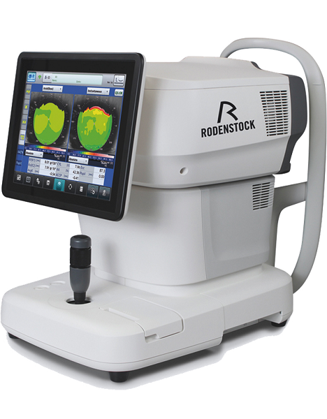
The Sequoia 512 with LCD ultrasound machine uniquely minimizes automatic image optimization time by up to 80% to make way for an easier and speedier ultrasound test time as well as allow for a more confident diagnosis each time. As a patient-specific imaging ultrasound, it has a unique matched response capacity for accurate image quality, sharp clinical specificity, and optimized diagnostic outcomes that makes infinitesimal pathologies and other anatomical detail in the most difficult-to-image patients becomes more vivid and visible. | Rodenstock Alino Multifunction Refractometer
Tune-up your businessExperience 6 analyses in 1 device: Rodenstock Alino Multifunction Refractometer
Are you ready for something that is all-in-one, timesaving, space-saving and suits your business perfectly?Have a look at how powerful ALINO can be.6 in 1 solution01 RefractionHaving a good starting value for subjective refraction is essential. The QUICK REF MODE supports this even for uncooperative patients.02 KeratometryKAI (Kerato-asymmetry index) & KRI (Kerato-regularity index) give the first hint of possible irregularities on the cornea. This supports evaluation of the best vision correction or best-fitting contact lens.03 TonometryThe very gentle air puff created by a new generation of air-flow technology means that patients more readily accept the need for regular check-ups of the IOP measurement.04 PachymetryCorrecting the IOP by measuring the central corneal thickness is essential in interpreting the intraocular pressure.05 TopographyVarious topographic maps support you in carrying out vision screenings, contact lens fittings and in patient education.06 Dry EyeObserve the customers’ eye health and evaluate the long-term tolerability of contact lenses.Take advantage of the combination of tests:Additional servicesTonometry + PachymetryContact lens fitting andtolerance controlKeratometry + Topography + Dry EyeEye healthcareTopography + Tonometry + Pachymetry + Dry EyeSpecifications Rodenstock Alino Multifunction Refractometer
REFRACTIVE POWER MEASUREMENTSpherical refractive power (S) -30.00 D to + 25.00 D (at VD = 12.0 mm)Cylindrical power (C) 0.00 D to ± 12.50 D (at VD = 12.0 mm)Astigmatic axis (A) 0° to 180°KERATOMETRY MEASUREMENTCorneal curvature radius 5.00 mm to 13.00 mmCorneal astigmatic axis 0° to 180°INTRAOCULAR PRESSURE MEASUREMENTMeasurement range 1 mmHg to 60 mmHG (1 hPa to 80 hPa)PACHYMETRY MEASUREMENTMeasurement range 300 μm to 800 μmTOPOGRAPHY MEASUREMENTCorneal curvature radius 5.50 mm to 10.00 mmCorneal astigmatic axis 0° to 180°AUXILIARY FUNCTIONInterpupillary distance Measurement range: 20 mm to 85 mmCorneal diameter and pupil diameter Measurement range: 1.00 mm to 14.00 mmDry eye observation Blinking Analysis, Tear Meniscus height, Hyperemia, Meibomian glandsDATA MANAGEMENTInternal Database Integrated SD CardPrinter Integrated thermal printerData Output Type 3x USB 2.0, 1x LAN, 1x SD Card-slot, 1x WLANExport Format CSV, XML, JPG, PDFDIMENSIONS & ELECTRIC REQUIREMENTSDimensions WDH 312 × 491 × 450 mmWeight Approx. 23 kgVoltage 100 VAC to 240 VACFrequency 50/60 HzPower consumption 110 VA | Nidek RS-330 Retina Scan Duo 2 with FAFThe Retina Scan Duo 2 is a combined OCT and fundus camera system that incorporates new features enhancing screening and clinical efficiency in addition to user-friendly features that were incorporated from the previous model. FEATURES : Nidek RS-330 Retina Scan Duo 2 with FAF- Fundus image acquisition with macula and disc capture in one image on OCT
- Combined diagnosis of macular and disc pathologies
- Wide area scan (12 x 9 mm)
Wide area normative database (macula: 9 x 9 mm, disc: 6 x 6 mm): A 12 x 9 mm wide area image can be acquired. The retina map captures both the macula and disc in a single shot. The normative database provides a wide area color-coded map comparing the patient’s macular thickness to a population of normal eyes.
- Denoising technique with deep learning: A new image enhancement technique using deep learning automatically displays a denoised image once B-scan acquisition is complete. With deep learning of a large data set of images averaged from 120 images, this denoising technique provides high definition images comparable to a multiple-image-averaging technique. The denoising function generates high definition images from a single frame while decreasing image acquisition time and increasing patient comfort.
- Quick acquisition of high definition B-scan images from a single-frame image
- Fundus autofluorescence (FAF): The FAF function is an advanced screening feature that allows non-invasive evaluation of the RPE without contrast dye.
*Available for the FAF model
Nidek RS-330 Retina Scan Duo 2– Easy operation with 3-D auto tracking, auto shot, and user friendly interface
– High definition images
– Wide area scan (12 x 9 mm) / Wide area normative database (9 x 9 mm)
– Multiple OCT scan patterns
– Value added features
– Various reports
OCT images are captured at scan speeds of 70,000
A-scans/s which is 32% faster than acquisition with the
Retina Scan Duo™ using Regular OCT sensitivity* Wide area scan (12 x 9 mm)
Wide area normative database (macula: 9 x 9 mm, disc: 6 x 6 mm)
A 12 x 9 mm wide area image can be acquired. The retina
map captures both the macula and disc in a single shot.
The normative database provides a wide area color-coded
map comparing the patient’s macular thickness to a
population of normal eyes. Denoising using deep learning
A new image enhancement technique using deep learning automatically displays a denoised image once
B-scan acquisition is complete. With deep learning of a large data set of images averaged from 120 images,
this denoising technique provides high definition images comparable to a multiple-image-averaging
technique. The denoising function generates high definition images from a single frame while decreasing
image acquisition time and increasing patient comfort | New ZEISS CIRRUS 6000 – 100 kHz OCT Imaging SystemAt 100,000 scans per second, ZEISS CIRRUS 6000 enables clinicians to image a larger field of view up to 12mm in a single scan. It also captures high-defintion (HD) OCT and OCT Angiography (OCTA) scans, revealing the finer microvascular details of the retina and providing more insight into your patient’s condition. AngioPlexOCT Angiography from ZEISS ushers in a new era of eye care with non-invasive imaging of retinal microvasculature—taking glaucoma and retinal disease management and treatment planning to the next level. By offering the industry’s most comprehensive tools for assessing and analyzing a range of pathologies, ZEISS provides a complete OCT Angiography (OCTA) solution. CIRRUS 6000 is the latest addition to an already robust portfolio of OCT devices that are designed to meet the needs of all types and levels of practices and/or clinicians. It offers customers high-speed and high-throughput to help manage their large patient volume faster, all while improving imaging and image quality. “This system was designed with the patient in mind, enabling doctors to continue accessing their patient data from CIRRUS devices that are crucial to effective long-term patient care,” said Jim Mazzo, Global President of Ophthalmic Devices at Carl Zeiss Meditec. “We are committed to our customers and believe this will become an essential asset to their practice.”The 100kHz speed of the CIRRUS 6000 allows clinicians to scan patients faster, with increased efficiency and improved imaging detail. Additional features include: - Wider and deeper OCT/OCTA scans with 12x12mm OCTA and B-scan depth up to 2.9mm
- HD AngioPlex scan is a new scan type that provides more detail
- New workflow protocols to aid in increasing efficiency
- Patient-friendly “Wellness report” to help educate the patient
According to retina and vitreoretinal physician and surgeon Gregg T. Kokame, MD, of Retina Consultants of Hawaii, the new ZEISS CIRRUS 6000 is extremely fast and impressive. “With the new scan acquisition time of 100,000 per second I can scan and assess my patients much quicker, giving me the details I need with every image,” says Kokame.“ZEISS is dedicated to advancing eye care through development of new and innovative solutions, like the CIRRUS 6000,” said Dr. Ludwin Monz, President and CEO of Carl Zeiss Meditec AG. “With our comprehensive diagnostics portfolio, we will continue to create better vision standards to benefit patients today and in the future.” | Nidek OPD-Scan III Corneal AnalyzerComprehensive vision analysis
OPD-Scan III VS is a device that mainly measures corneal shape (topographer) and refractive error (refractometer).
It clarifies causes of vision difficulties through wavefront analysis of information measured over a wide area.
Solutions for face-to-face consultingTablet viewer provides four kinds of easy-to-understand reports for explanation and consulting.
*The tablet is not included or sold with the OPD-Scan III VS.
Basic information reportBasic information to generally judge the patient’s eye conditions.
Topography reportIntuitive maps and numerical data for comprehending the corneal surface.
Simulation reportVisual performance simulations and MTF graphs for a variety of conditions.
Eye diagram image reportEye model to visually understand eye conditions ranging from eye fundus to cornea.
Wide range of display patterns
  Placido image
 Axial map (Additional indication)
 Visual Acuity map (Image charts)
Features Nidek OPD-Scan III Corneal Analyzer- Five-in-One true refractive workstation for all practitioners
- Overview summary for optimal clinical decisions
- A number of summaries
- Wider measurement area
- Greater topography resolution, blue placido rings
The OPD-Scan lll is the Five-in-One true refractive workstation combining - Wavefront Aberrometer
- Topographer
- Auto Refractometer
- Auto Keratometer
- Pupillometer and Pupillographer
- Irregularity helps determine the best strategy for vision correction. Separation into Total, Corneal and Internal components allows determination of the source of the optical pathology.
- PSF images of OPD, Axial, and Internal OPD map simulate objective retinal visual quality from each component of the eye for easy clinical assessment and patient education.
- Corneal Spherical Aberration aids in the selection of aspheric IOLs and contact lenses.
- Color coded Classification Indices help identify post-LASIK corneas and Keratoconus.
- The Astigmatism index aids the implantation of toric IOLs such as incision placement and lens alignment.
- A retroillumination image of cataracts captured during the OPD exam allows better understanding of pupillary effects on vision and in patient education.
|




Reviews
There are no reviews yet.