View cart “Sonosite M-Turbo Ultrasound Machine For Sale” has been added to your cart.
-50%
Availability: In Stock
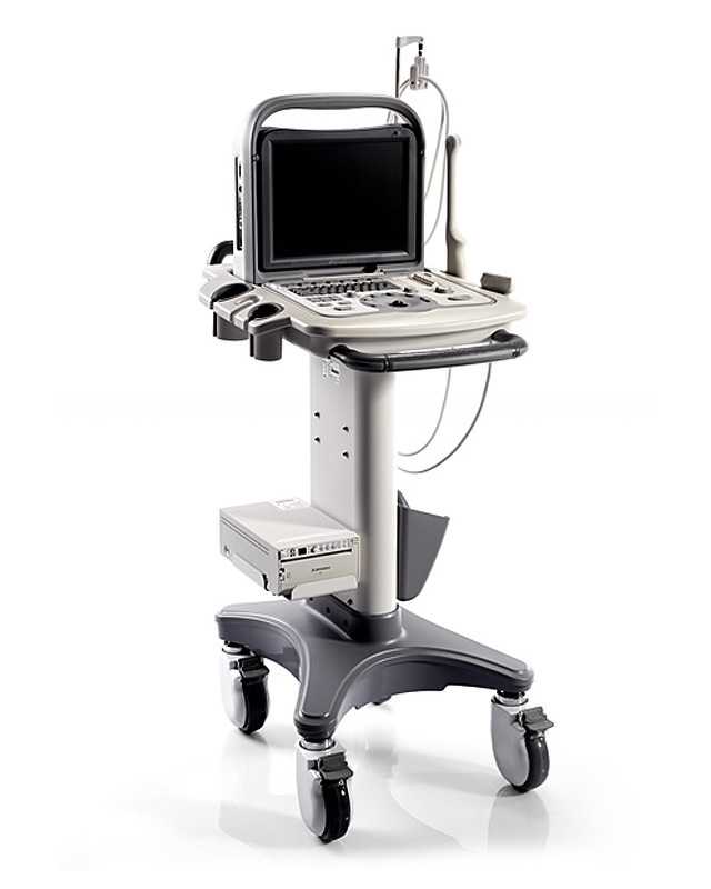
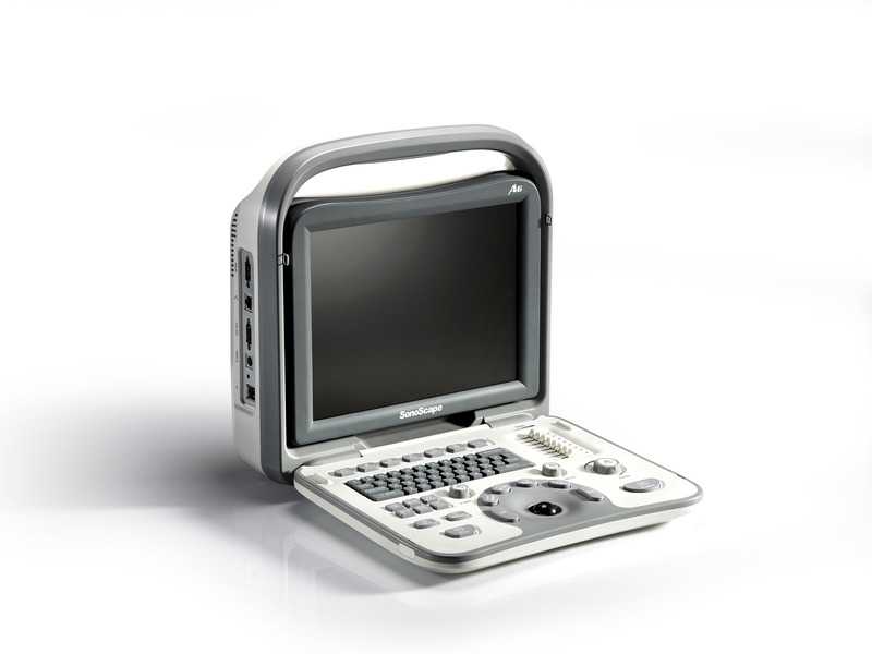
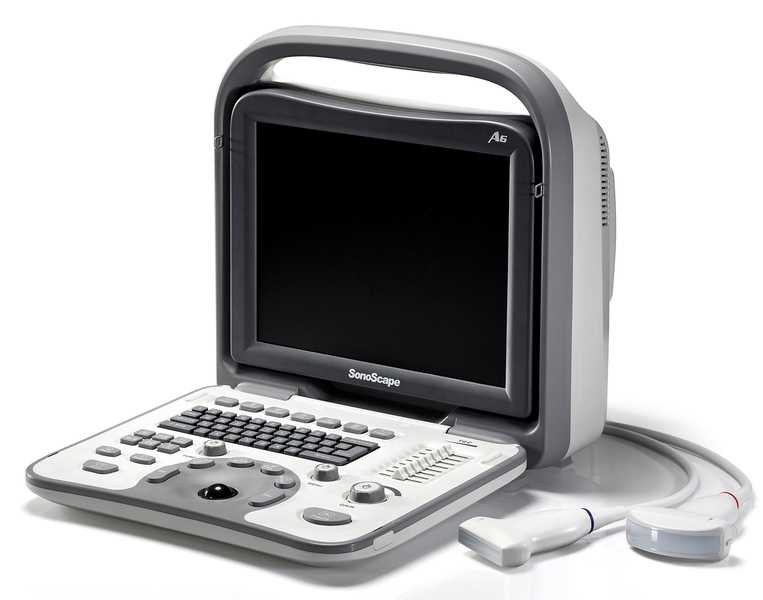
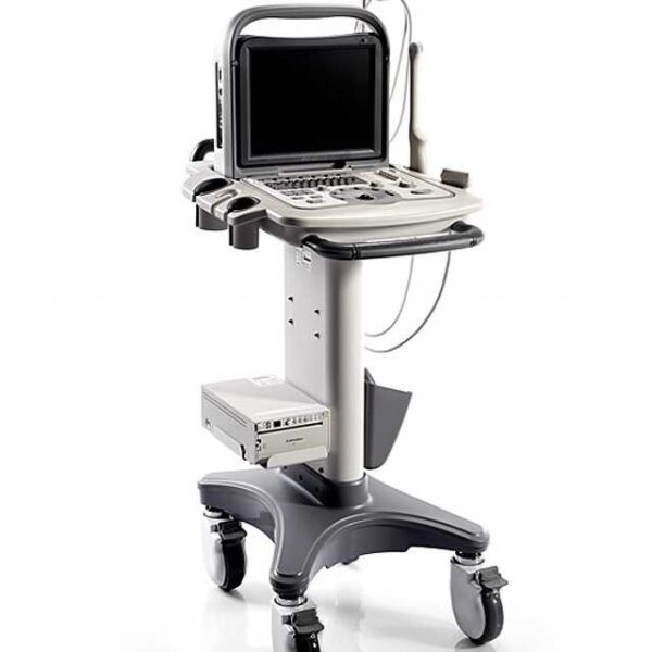
SonoScape A6 Portable Ultrasound System For Sale
$7,000.00$3,500.00
SonoScape A6 is a low-cost black system. Two active probe interfaces are available and internal battery allows to scan for up to 3 hours on a complete charge.
Other people want this. 15 people have this in their carts right now.
Reliable and easy to use hand-carried ultrasound system
SonoScape A6 is a mobile ultrasound system considered best among white and black imaging ultrasound machines. It’s frequency range around 12MHz on its own linear probes and is a great choice for general imaging, line positioning, OB, MSK, and needle advice. It’s a cost-effective choice for physicians in need of a high-resolution B/W machine for their clinic.
SonoScape A6 is concentrated and dependable equipment for clinics and hospitals. A6 was started to meet the requirements of both doctors and patients. Sonoscape A6 is your all-around greatest B/W ultrasound machine and yet stays cheap.
The Sonoscape A6 portable black and white ultrasound equipment is a popular ultrasound used for OBGYN, vascular, liver, muscle-skeletal and other sorts of studies. The ultrasound has simple to use navigation using simple to reach study configurations and applications.
SonoScape A6 includes a deeper scanning thickness, Bi-Plane transducer option, chroma function, greater zoom, more streamlined, LCD screen and better imaging. It is very helpful in diagnosing liver, kidney, abdomen, biopsies, OB/GYN, vascular access and boat advice. It’s full patient database solutions such as DVD, USB storage, PDF report, AVI/JPEG, DICOM3.0, etc..
Application:
Ob/Gyn
Anesthesia
Urology
Vascular
Access
MSK
Breast Imaging
Small Parts
Features:
12” Adjustable LCD Display
Fully Digital Beamformer
Two Probe Connectors
Tissue Harmonic Imaging
B-Mode
M-Mode
Panoramic Imaging
Zoom
Automatic Image Optimization
DICOM 3.0
Options:
Mobile Trolley
External DVD RW
Carry Case
Printer
System Specifications:
Image/Video Formats: BMP, JPG, and AVI Video
Storage Types: USB, DVD, Hard Drive VGA, S-Video, Print Output
SonoScape A6 is a low-cost black system. Two active probe interfaces are available and internal battery allows to scan for up to 3 hours on a complete charge.
The more patients you see, the more you need Voluson E8.
The Voluson E8 ultrasound system is designed to keep pace with busy practices that conduct a wide range of Women’s Health exams, from routine scanning to complex assessments.
The imaging excellence of Radiance System Architecture – your diagnostic confidence will be enhanced by the extraordinary image clarity and speed of Radiant System Architecture.
Designed to make your job easier, faster and more accurate, the MicroMaxx ultrasound tool makes point-of-care ultrasound an affordable reality. It offers impressive image quality. Wireless data transfer. Extreme durability. The Sonosite Micromaxx also features SonoCalc IMT, edge detection software for cardiology and Tissue Harmonic Imaging
The Samsung Accuvix XG was the top of the line system from Samsung prior to the release of the newer. The XG was meant as a direct replacement to the popular Accuvix XQ as a shared service system capable of both 4D OBGYN images as well as full cardiac scans.
Acuson Cypress cardiovascular system PLUS delivers advanced ergonomics and superior performance in a highly portable package. The Cypress System PLUS is a highly miniaturized, all digital, phased array echocardiography system that provides complete studies and outstanding images – even on patients who are difficult to image.
The SonoSite Titan is the earliest version of the PoC dedicated portable ultrasound machine from Sonosite. Though its form factor is compact, it provides high resolution imaging, rapid responsive time, and supports a broad range of applications including cardiac.
Content
SonoScape A6 Portable Ultrasound System
Reliable and easy to use hand-carried ultrasound systemSonoScape A6 is a mobile ultrasound system considered best among white and black imaging ultrasound machines. It’s frequency range around 12MHz on its own linear probes and is a great choice for general imaging, line positioning, OB, MSK, and needle advice. It’s a cost-effective choice for physicians in need of a high-resolution B/W machine for their clinic.SonoScape A6 is concentrated and dependable equipment for clinics and hospitals. A6 was started to meet the requirements of both doctors and patients. Sonoscape A6 is your all-around greatest B/W ultrasound machine and yet stays cheap.
The Sonoscape A6 portable black and white ultrasound equipment is a popular ultrasound used for OBGYN, vascular, liver, muscle-skeletal and other sorts of studies. The ultrasound has simple to use navigation using simple to reach study configurations and applications.SonoScape A6 includes a deeper scanning thickness, Bi-Plane transducer option, chroma function, greater zoom, more streamlined, LCD screen and better imaging. It is very helpful in diagnosing liver, kidney, abdomen, biopsies, OB/GYN, vascular access and boat advice. It’s full patient database solutions such as DVD, USB storage, PDF report, AVI/JPEG, DICOM3.0, etc..Application:
Ob/Gyn
Anesthesia
Urology
Vascular
Access
MSK
Breast Imaging
Small Parts
Features:
12” Adjustable LCD Display
Fully Digital Beamformer
Two Probe Connectors
Tissue Harmonic Imaging
B-Mode
M-Mode
Panoramic Imaging
Zoom
Automatic Image Optimization
DICOM 3.0
Options:
Mobile Trolley
External DVD RW
Carry Case
Printer
System Specifications:
Image/Video Formats: BMP, JPG, and AVI Video
Storage Types: USB, DVD, Hard Drive VGA, S-Video, Print Output
As your patient volume increases and your clinical cases become more complex, you will require an ultrasound system that enables you to deliver truly exceptional care – confidently and efficiently – every time. The Voluson E8 ultrasound system is designed to keep pace with busy practices – handling routine to complex women’s health exams with ease and precision. The premium image quality of the Voluson E8 combined with access to advanced capabilities, delivers the level of performance required by you and your patients.
SonoSite MicroMaxx Ultrasound Machine
The SonoSite MicroMaxx Portable Ultrasound is the third portable system to emerge from SonoSite. With improved image quality, a larger screen and additional imaging features, this system is the upgrade to the SonoSite Titan. The SonoSite MicroMaxx portable ultrasound is a well suited portable ultrasound, and is capable of performing the duties of a shared service system.The portable SonoSite MicroMaxx Portable Ultrasound system has purpose-built software and proprietary ASIC design yielding high-quality images with speed and reliability while efficiently operating on either AC or battery power. From busy offices and big-city hospitals to critical-care situations where seconds count, the handheld SonoSite MicroMaxx ultrasound system, boots up quickly, from off to scanning in under 15 seconds. Sonosite Micromaxx portable ultrasound transducers, probes, service and pricing are available through UDS Portable Ultrasound.Big machine performance in a hand-carried unitMicroMaxx’s purpose-built software and proprietary ASIC design yields high-quality images with speed and reliability while efficiently operating on either AC or battery power.MicroMaxx goes anywhere you need itFrom busy offices and big-city hospitals to critical-care situations where seconds count, the SonoSite MicroMaxx&trade system boots up quickly, from “off” to scanning in under 15 seconds. This easily carried device gets medical professionals into-and out of-tight spaces fast.Embedded software means solid reliabilityThe MicroMaxx system software is hard-wired and purpose-built for faster boot.Chip Fusion TechnologySonoSite’s proprietary Chip Fusion™ Technology enables advanced, cart-based image resolution in a hand-carried unit by integrating digital signal processing and multiple ultrasound functions into a custom ASIC microchip to deliver unprecedented performance without the size, weight and cost of conventional systems.Clinical utilityThe MicroMaxx system comes with a wide range of transducers for multiple clinical applications. With MicroMaxx, SonoSite is extending the capabilities of hand-carried systems by adding high-performance features.Durability SonoSite MicroMaxx Ultrasound MachineSonoSite products and technology deliver proven reliability and durability in conventional medical settings as well as some far more challenging environments, such as supporting rescue operations following natural disasters. Extensive quality controls such as “drop testing” ensure that SonoSite products continue to set the industry standard for both reliability and durability.MicroMaxx’s reliability and durability are backed by a one-year warranty, an industry first, far exceeding warranties available on competing products.From “Off” to scanning in just secondsThis is vitally important in many medical situations where seconds really do count-whether getting crucial diagnostic information in an emergency or making the best use of time in a hectic daily schedule.
Samsung Accuvix XG Ultrasound Machine2D Image Features
• Dynamic MR™ / Dynamic MR Plus is designed to enrich gray-scale resolution, as it enhances detection
and contrast resolution while also decreasing speckle echoes. This is particularly useful when evaluating
superficial structures, including thyroid, vessels, pelvic and abdominal anatomy.
• SRF (Speckle Reduction Filter™) enhances image quality by reducing or eliminating the appearance of
speckle echoes from ultrasound images. The degree of speckle reduction implemented is user-selectable.
• Wide Dynamic Range determines the number of gray shades utilized to map the gray-scale image.
It enables display of more details of bright areas and dark areas.
• SCI (Spatial Compounding Image) controls the ultrasound beam electronically by steering, and compounds
many scan lines.
• FSI (Full Spectrum Imaging) incorporates the penetration capabilities associated with lower frequencies,
yet maintains the fine pixel uniformity associated with higher frequencies, to deliver consistently high quality
images even in challenging diagnostic areas.3D Image Features Medison Accuvix XG Ultrasound System
• HDVI gives outstanding image quality and naturally clearer contrast, with excellent tissue differentiation,
edge depiction and speckle reduction, allowing consistent diagnoses with great confidence.
• 3D XI™, comprised of a suite of three innovative imaging applications – Multi-Slice View, Oblique View™ and
Volume CT – offers complete and precise control over 3D/4D volume data manipulation for maximum
diagnostic accuracy.
• 3D MXI is an innovative, cutting-edge 3D image processing technology. Comprising a comprehensive suite of
imaging tools – including Multi Volume Slice, Mirror View, Multi-OVIX, and 3D OH – 3D MXI lets you view,
examine and diagnose 3D volume data with supreme ease, speed and accuracy. • Fully adjustable system
The control panel can be adjusted to the user’s preferred height, for a better working environment and reduced
risk of back pain.
• Wide LED touch-screen
The Accuvix XG’s new LED touch-screen makes it easy to organize and operate.
• 19-inch HD LCD Monitor and articulating monitor arm
A 19-inch LCD monitor enables images to be displayed clearly even with a larger monitor, and the articulating
monitor arm enables easy mobility for a more comfortable and convenient working environment.
• Portability
The Accuvix XG is a lightweight system with 4 swivel wheels that allow easy steering, and a locking function. • Customizable measurement menus
Customizable measurement menus allow access to frequently-used functions, and enable a quicker and
more intuitive workflow.
• User keys and user knob
Accuvix XG offers user keys and a user knob that can map frequently-used functions, enabling the function to be
activated quickly and easily.
• Customized Annotation Menu and body marker
Users can preset up to 360 words of annotations, and body markers for each application, that reduce the time
needed for each examination.
• QuickScan
QuickScan maximizes workflow efficiency by automatically optimizing key imaging parameters with just the
push of a button. • HDVI
HD Volume Imaging(HDVI) gives outstanding image quality and naturally clearer contrast, with excellent tissue
differentiation, edge depiction and speckle reduction, allowing consistent diagnoses with great confidence.
It shows clearer images of subtle lesions, using multi slice image as well as C-plane.
• ElastoScan
ElastoScan™ translates stiff & soft tissue information into gray scale image. This technology defines the alteration
of the stiffness in tissue state more accurately, making it easier for the user to utilize the information in the image.
Acuson Cypress PLUS Ultrasound
Acuson Cypress cardiovascular system PLUS delivers advanced ergonomics and superior performance in a highly portable package. The Cypress System PLUS is a highly miniaturized, all digital, phased array echocardiography system that provides complete studies and outstanding images – even on patients who are difficult to image.The Cypress PLUS transitions easily from your small private office or hospital echo lab to the operating theatre or your patient’s bedside. It’s ultrasound where you need it.TheCypress PLUS delivers high performance in a complete range of cardiovascular capabilities.Designed with advanced ergonomics in mind, the Cypress system PLUS streamlines your workflow and increases your productivity.If you already own a Cypress system, keeping technologically current is simple with the Cypress Upgrade program.The Cypress system is the smallest, lightest, high performance, phased array echo system ever built. Weighing in at just 19 pounds, with digital storage capacity for over 100 studies and network capabilities to send studies from remote locations, clinicians can perform complete echocardiography studies from almost anywhere. The Cypress system was designed for portability, durability and reliability, and to withstand the rigors of constant transportation. Internal system interconnects are minimized. Key components are shock mounted and the entire system sits inside a robust chassis. From hospital to office or from airplane to mountain village, the Cypress system will perform.Full Range of Capabilities
TheCypress system is a complete, full-featured echo cardiography system that provides complete studies and outstanding image quality even on the most difficult to image patients. It offers a full range of features including:– 2D with Harmonics
– Patient Report with audio capture
– M-Mode
– Embedded DICOM conformance
– Color PW and CW Doppler
– High frame rate color flow imaging
– Stress echo
– Network output
– Vascular imaging
– Comprehensive calculations package
– Contrast agent imaging
– USB peripheral support (PC printer connectivity)Physical Characteristics Acuson Cypress PLUS Ultrasound• Single unit houses ultrasound system with control panel, flat panel display and MO drive
• Weight: 8.6 Kg (19 pounds)
• Height: 35.6 cm (14 in)
• Width: 40.6 cm (16 in)
• Depth: 20.3 cm (8 in) – with keyboard folded up 34.3 cm (13.5 in) – with keyboard down
• Heat dissipation: 490 BTU/hour (nominal)User Interface
• Fold-up control panel for portability
• Direct access to system functions through dedicated keyboard controls and multifunctional soft keys
• Easy-access main controls, including multifunctional trackball, enter and select keys, for minimum hand movement while controlling the system
• Adjustable trackball speed
• Functional grouping of keys, rotational knobs, switches and sliding TGC controls for direct access to critical functions
• Tactile feedback on control activation
• Backlighting of control panel labels
• Mode-dependent soft keys
• User-interface tools
• Pre-defined and user-entered labeling textMonitor
• 10.4 inch/26.4 cm flat panel active matrix display screen
• Protective anti-glare, anti-reflective lensHard Drive
• Internal hard drive for image and data storage
• Internal storage of approximately 50 patient studies (25 – 30 loops per study)Transducer Ports
• One transducer connector (type IEC601BF)Operating/Display Modes
• 2-D, fundamental and harmonic imaging
• Color Doppler Velocity (CDV)
• Color Doppler Energy (CDE)
• M-mode, vertical split screen display with 2-D
• Pulsed Wave (PW) Spectral Doppler
• Continuous Wave (CW) Spectral Doppler
• Duplex Doppler (combined 2-D and Spectral Doppler display)2D Calipers – Generic Measurements and Calculations
• Measurements and calculations in 2D, M-mode, color Doppler, PW and CW spectral Doppler modes
• Basic measurements and report calculations
• Comprehensive patient calculations report
• On-screen “mini-reports”
• Complete or configured patient report
• Cardiac package
• Carotid package
• Arterial package
• Venous package
• Abdominal package
• User configurable Sites
• Selectable menu position
• Four cursor types
• Angle correction
• Distance
• Area
• Slope
• Time
• Velocity
• Velocity/pressure
• Mean velocity/pressure gradient
• Velocity Time Integral
• Pressure halftime
• Continuity Equation
• Ejection Fraction
• Left Ventricular Volumes, including End-Systolic and End-Diastolic volumes
• Cardiac output
• Cardiac index
• Fractional area changeCine Review
• 2D Cine memory up to 15 seconds of history
• Color Doppler cine memory up to 15 seconds of history
• M-mode strip cine along with PW and CW Doppler mode
• strip cine memory determined by sweep speed (selectable by the user) 10, 20, 30 or 40 secondsTransducer Technology
• 3V2c: 2-4 MHz wideband phased array
• 7V3c: 3-7 MHz wideband phased array
• 7L3: 5-7 MHz wideband linear array (Vascular)
• 4C1: 2-4 MHz wideband curved linear array (Abdominal)
• V5Ms: 3-6 MHz wideband phased array (micro-pinless (MP) connector)
Sonosite Titan Ultrasound Machine
The SonoSite Titan Portable Ultrasound is SonoSite’s second generation portable ultrasound system. This system improved upon the technology of the SonoSite 180 with a larger screen and keyboard packed into a laptop style system. This system is well suited for Vascular access, Musculoskeletal, Anesthesiology, Internal Medecine, Emergency rooms and Family Physicians. Veterinary configurations are available. Veterinary configurations are available.8.4″ LCD monitor, 2 probe ports, Vascular, OB/GYN, Cardiac reports, Soft keys, Split screen, Zoom, Storage via 64 or 256 MB flash cards, 220 image CINE loop, USB & Ethernet image transfer, Volume calculations via diameters, THI [Tissue Harmonic Imaging], SonoCalc [IMT analysis, PC-based], DICOM, S-video (in/out), RGB, Composite video.
Dimensions: 3″ (H) x 10.9″ (W) x 11.9″ (D) Weight: 7.7 lbs
C60 – abdominal/ obstetrics/ gynecology – 5-2 MHz 60-mm broadband curved array – 22 cm – Available – TITAN
ICTe – obstetrics/ gynecology – 8-5 MHz 11-mm broadband tightly curved array – 10 cm – Available – MicroMaxx/ TITAN
L25 – nerve/ musculoskeletal/ vascular and superficial – 10-5 MHz 25-mm broadband linear array – 6 cm – Transverse Guide Available – TITAN
L38 – small parts/ breast/ vascular/ nerve/ musculoskeletal/ superficial – 10-5 MHz 38-mm broadband linear array – 6.5 cm – Available – TITAN
Options
Extended cardiac package
Pulsed wave (PW) doppler
Continuous wave (CW) doppler
Tissue Harmonic Imaging
Accessories
Cart
Black and White Video Printer
DVD Recorder
The SonoSite Titan is the earliest version of the PoC dedicated portable ultrasound machine from Sonosite. Though its form factor is compact, it provides high resolution imaging, rapid responsive time, and supports a broad range of applications including cardiac.



Reviews
There are no reviews yet.