View cart “GE Vivid Q Ultrasound Machine For Sale” has been added to your cart.
-43%
Availability: In Stock
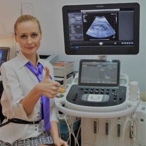
Philips EPIQ 5 Ultrasound Machine For Sale
$28,000.00$15,890.00
Philips EPIQ 5 might be the new leadership for a premium ultrasound, including an uncompromised level of clinical operation to meet the challenges of the toughest practices and technically difficult-to-image patients through each gestational age and for gynecology programs.
Other people want this. 15 people have this in their carts right now.
EPIQ 5 is the new direction for the superior ultrasound, featuring an uncompromised level of clinical operation to meet the challenges of the toughest general imaging practices.
The Philips Epiq 5 ultrasound system brings together the newest technologies using the ergonomics you need all in one system. The Epiq series offers a user-friendly interface which focuses on saving the operator time whilst maximizing workflow. Technologies like Epiq’s PureWave transducers coupled with precision technology delivers exceptional picture quality and the confidence to make an informed diagnosis.
It offers Purewave single crystal probes, a compact and lightweight design and superior design and workflow. The Epiq 5 replaces the elderly Philips iU22 with no xMatrix. The Epiq 5 has fewer probe choices and features compared to expensive Epiq 7. All the Epiq and Affiniti ultrasound machines look visually quite similar and use the exact same track and enormous tablet-like touchscreen for navigation.
Philips nSIGHT Imaging is an entirely new approach The Philips proprietary nSIGHT Imaging architecture introduces a totally fresh method of forming ultrasound images without compromise. Unlike traditional systems that form the image line by line, nSIGHT creates images with optimal resolution down to the pixel level.
Outstanding architecture Proprietary nSIGHT Imaging integrates the use of a brand new precision beamformer together with powerful massive parallel processing. This incredible structure captures an enormous Number of acoustic information and reconstructs optimally concentrated beams, creating a precise resolution for every pixel in the picture — all in real time
Designed for the user experience
EPIQ 5 is among the greenest systems we have ever designed. It consumes 25% less power compared to our current premium ultrasound. EPIQ 5 is almost quiet when running. A noise test determined that EPIQ 5 runs at 37-41 dB, which is equal to the sound of a library
Over 80% of sonographers experience work-related pain, and over 20% of them suffer a career-ending injury. With EPIQ 5 a new tablet-like interface results in dramatic reduction in reach and button pushes, with40% to 80% less reach and 15% fewer steps.
PHILIPS EPIQ 5 Ultrasound for Radiology with 2 Probe Premium. Three major settings such as Cardiovascular, Radiology and Common service / General Imaging.
Philips EPIQ 5 Features
The Philips Epiq 5 ultrasound system brings together the newest technologies with the ergonomics you want all in one system. The Epiq series offers a user-friendly interface that focuses on saving the operator time while maximizing workflow. Technologies like Epiq’s PureWave transducers coupled with precision engineering delivers exceptional image quality and the confidence to make an informed diagnosis.
Lightweight: 230 lbs
Highly ergonomic
12” Tablet-like touchscreen user interface
auto doppler
High-resolution 21.5” LCD widescreen monitor
4 probe connectors with fast switch capability
PureWave-Crystal-Technology
nSIGHT: clear, precise imaging throughout the image while keeping higher framerates
SmartExam & Real Time iSCAN (Autoscan)
PureWave transducers
4D TEE
Shear Wave and strain-based elastography
2D LV quantification post-scan aiming guidance to minimize uncertainty
Off-center scan and pubic bone interference notifications
Intuitive user interface
Onboard video tutorial for training and ease of use
Customizable savings calculator to track the cost of ownership
Philips EPIQ 5 might be the new leadership for a premium ultrasound, including an uncompromised level of clinical operation to meet the challenges of the toughest practices and technically difficult-to-image patients through each gestational age and for gynecology programs.
The GE Vivid q portable ultrasound machine is a more powerful version of the Vivid i that adds further quantitative features including ICE, Auto EF, AFI, TVI,TT, TSI, Blood Flow and Blood Flow imaging. The Vivid q is the pinnacle of GE’s portable cardiovascular ultrasounds.
Function • Gravity Sensor: You can operate the ultrasound system either in transversal layout or vertical layout.• Grid for estimation: With grid on the image area, you can easily estimate the size of the target substance like follicle fertilized egg etc.
MedCorp is a trusted source for new and refurbished ultrasound systems with a combined 50 years experience in the industry and a detailed process for ensuring that each system they sell is as good as the day it was manufactured.
Acuson Cypress cardiovascular system PLUS delivers advanced ergonomics and superior performance in a highly portable package. TheCypress System PLUSis a highly miniaturized, all digital, phased array echocardiography system that provides complete studies and outstanding images – even on patients who are difficult to image.
The Samsung Accuvix XG was the top of the line system from Samsung prior to the release of the newer. The XG was meant as a direct replacement to the popular Accuvix XQ as a shared service system capable of both 4D OBGYN images as well as full cardiac scans.
Content
Philips EPIQ 5 Ultrasound Machine
Most powerful premium ultrasoundEPIQ 5 is the new direction for the superior ultrasound, featuring an uncompromised level of clinical operation to meet the challenges of the toughest general imaging practices.The Philips Epiq 5 ultrasound system brings together the newest technologies using the ergonomics you need all in one system. The Epiq series offers a user-friendly interface which focuses on saving the operator time whilst maximizing workflow. Technologies like Epiq’s PureWave transducers coupled with precision technology delivers exceptional picture quality and the confidence to make an informed diagnosis.
It offers Purewave single crystal probes, a compact and lightweight design and superior design and workflow. The Epiq 5 replaces the elderly Philips iU22 with no xMatrix. The Epiq 5 has fewer probe choices and features compared to expensive Epiq 7. All the Epiq and Affiniti ultrasound machines look visually quite similar and use the exact same track and enormous tablet-like touchscreen for navigation.Philips nSIGHT Imaging is an entirely new approach The Philips proprietary nSIGHT Imaging architecture introduces a totally fresh method of forming ultrasound images without compromise. Unlike traditional systems that form the image line by line, nSIGHT creates images with optimal resolution down to the pixel level.Outstanding architecture Proprietary nSIGHT Imaging integrates the use of a brand new precision beamformer together with powerful massive parallel processing. This incredible structure captures an enormous Number of acoustic information and reconstructs optimally concentrated beams, creating a precise resolution for every pixel in the picture — all in real timeDesigned for the user experienceEPIQ 5 is among the greenest systems we have ever designed. It consumes 25% less power compared to our current premium ultrasound. EPIQ 5 is almost quiet when running. A noise test determined that EPIQ 5 runs at 37-41 dB, which is equal to the sound of a libraryOver 80% of sonographers experience work-related pain, and over 20% of them suffer a career-ending injury. With EPIQ 5 a new tablet-like interface results in dramatic reduction in reach and button pushes, with40% to 80% less reach and 15% fewer steps.PHILIPS EPIQ 5 Ultrasound for Radiology with 2 Probe Premium. Three major settings such as Cardiovascular, Radiology and Common service / General Imaging.Philips EPIQ 5 FeaturesThe Philips Epiq 5 ultrasound system brings together the newest technologies with the ergonomics you want all in one system. The Epiq series offers a user-friendly interface that focuses on saving the operator time while maximizing workflow. Technologies like Epiq’s PureWave transducers coupled with precision engineering delivers exceptional image quality and the confidence to make an informed diagnosis.
Lightweight: 230 lbs
Highly ergonomic
12” Tablet-like touchscreen user interface
auto doppler
High-resolution 21.5” LCD widescreen monitor
4 probe connectors with fast switch capability
PureWave-Crystal-Technology
nSIGHT: clear, precise imaging throughout the image while keeping higher framerates
SmartExam & Real Time iSCAN (Autoscan)
PureWave transducers
4D TEE
Shear Wave and strain-based elastography
2D LV quantification post-scan aiming guidance to minimize uncertainty
Off-center scan and pubic bone interference notifications
Intuitive user interface
Onboard video tutorial for training and ease of use
Customizable savings calculator to track the cost of ownership
The powerful GE Vivid Q ultrasound machine is the ultimate lightweight, compact high-performance cardiovascular system for clinicians needing a machine that combines user efficiency and reliable performance. This unique system performs consistently thanks to its TruScan architecture and can support up to 17 probes. The GE Vivid Q is recognized for its premium image quality, advanced image management, and high-performance quantification tools.This advanced GE ultrasound delivers improved patient care and enhanced user productivity with the new M4s-Matrix array technology transducer, which provides greatly enhanced image quality in electronic beamforming, raw data image acquisition and archiving, optimal onboard and post processing technology.This ultrasound system provides high clinical values for quantification and review of stored images, making it one of today’s most sought-after models. Some of its powerful features include compatibility with EchoPac clinical workstation and DICOM networking, speckle tracking strain, integrated stress echo, quantification tools, and reporting capabilities. Also of note is one of the Vivid Q’s best new features, which is its TEE transducer capability.A one-touch button optimization provides premium image quality for workflow efficiency, and maintains AFI- automatic function imaging with complete wallFeatures
Advanced QScan Imaging (incl TSI)
AdvStress
ATO
Auto EF
Automated Function Imaging
Blood Flow Imaging
CardiacApp
Cardio Lab
Color Doppler
CW Doppler
DICOM Networking
DICOM: Print
DICOM: Worklist
DVD/RW
ECG
ECG Cable
EchoDicom
EchoPAC
Evue
ICE Imaging
Intima Media Thickness (IMT)
LVO Contrast
M Mode
M4S
MPEGvue
PW Doppler
Small Depth
TEE
THI
Tissue Harmonics Imaging (THI/NTHI)
Tissue Velocity Imagine & Tissue Tracking
TSI
VascAbdContrast
VascularApp
VirtualPrinter
Wide Aperture
GE Vivid Q FeaturesThe GE Vivid Qis a Portable Laptop-style Ultrasound that can be used to help diagnose cardiovascular anatomy. Features and tools based on the previous Vivid 7 and the Vivid E9 have been added to the new lightweight Vivid Q, mainly the M4S-RS transducer that allows for 3D image focusing and enables a higher level of image quality. The Vivid Q ultrasound is versatile and can additionally be used for Cardiac, vascular, Abdominal, Pediatric, Obstetrics, and Emergency medicine. The Vivid Q Ultrasound comes with a 15-inch color LCD monitor that can display a variety of imaging modes including; ICE, Auto EF, AFI, TVI, TT, TSI, and Blood Flow. Additional the Ultrasound system is comparable with 17 different transducers and probes to fit every imaging need.
Auto EF - Automated ejection fraction measurement program designed specifically for the GE Vivid q.
Tissue Synchronization Imaging (TSI).
Intracardiac Echo (ICE).
16 different compatible probes.
Coded Octave imaging - enhances image color flow and Doppler imaging.
SIUI CTS-800 Veterinary Ultrasound Machine with Linear Rectal Probe
This small and convenient “Palm-Sized” portable veterinary ultrasound scanner fits in the palm of your hand, and is mostly used for farm animals. Its long battery life and waterproof features makes it the perfect ultrasound scanner for veterinarians on the field.The screen of the SIUI CTS-800V will rotate horizontally or vertically, according to how you are holding it.Gravity Sensor: You can operate the ultrasound system either in transversal layout or vertical layout. It can automatically adapt to two different layouts when you rotate it.Grid for estimation: With grid on the image area you can easily estimate the size of the target substance like follicle fertilized egg etc.SPECIFICATIONS SIUI CTS-800 Veterinary Ultrasound Machine with Linear Rectal ProbeMonitor: 7-inch WVGA LCD monitor
– Main unit with 7-inch LCD monitor.
– 1 Probe connector.
– 1 Li-ion battery. 3 hours operation time.
– 1 Adaptor.
– 1 Car Charger.
– Belt and sun shield.
– 1 USB port (Mini).
– Carrying case.PROBES– Rectal Linear L7FVC (6.5/7.1/7.5/8.3/9.0MHz).
– Rectal Linear L5FVC (4.0/4.7/5.0/5.5/6.2MHz).
– Backfat Linear L1FC (150mm, 2.5/3.0/3.5/4.0/5.0MHz).
– Convex C3FC (2.5/3.0/3.5/4.0/5.0MHz).
– Microconvex C5FC (3.5/4.0/5.0/6.5/7.1MHz).CTS-800 is a palm-sized veterinary ultrasound system. Features Weight: 0.8kg Environmental rating: IP54 (main unit) IP67 (probe head) Battery: 3 hours operation time Display mode: B 2B ZOOM B B/M M Depth: Max to 250mm B gain: 0-100 Image storage: 256 frames Cine loop: 256 frames Probe: L50/7.5 MHz linear rectal probe (standard) L65/5.0 MHz linear rectal probe (optional) R20/5.0 MHz micro convex probe (optional)
SonoSite Nanomaxx Ultrasound Machine
The SonoSite NanoMaxx ultrasound system combines premium functionality and affordability with the versatility and portability that makes SonoSite a top competitor in the portable ultrasound market. The SonoSite NanoMaxx comes in a sleek, hand-held package that never compromises performance, with applications ranging from emergency medicine and anesthesia to vascular.MedCorp offers the refurbished SonoSite NanoMaxx for purchase. Used SonoSite NanoMaxx from MedCorp offer everything you’d expect from a newer machine, including PC/MAC export configurations, 2D, Color and Color Power Doppler modes, and 2GB of internal memory that can hold 50 images each for 40 patients, all in an 8” touch-screen interface. However, because this is a refurbished SonoSite NanoMaxx, the price is lower than the new models.
Comes with:* NanoMaxx Ultrasound System, 1.0.1
* Battery – NanoMaxx 11.25 volt, 5.2 amp-hour rechargeable lithium-ion battery
* L38N / 10-5 MHZ Transducer multi-frequency, broadband, 38 mm linear array transducer for small parts, breast, vascular, nerve, IMT, and venous. Supports 2D with SonoMB Multi Beam Technology and Auto Gain capabilities, SonoHD Imaging Technology, Color Power Doppler, velocity Color Flow Doppler. Maximum depth is 9 cm.
* Power Supply, Universal supply (50-60 Hz, 100-240 VAC) for charging battery and powering the NanoMaxx.
* NanoMaxx Carry Case
8″ Touch-screen interface
2D, Color and Color Power Doppler modes
2GB internal memory that can hold 50 images each for 40 patients
2 hours of battery power
PC/MAC export configuration
Four preset pictograph screen positions
Drop resistant magnesium case
System Description:SonoSite Nanomaxx Ultrasound Machine
The NanoMaxx ultrasound system offers excellent performance at an affordable price. It is easy to use and offers top of the line diagnostic imaging, color power Doppler and color-flow velocity. The NanoMaxx can aid users in making clinical decisions or guide interventional procedures. The unit uses proprietary technology which allows the optimization of many system settings by simply pressing a button.Application:
Abdominal, Cardiology, Gynecology, Nerve, Obstetrics, CIMT, Musculoskeletal, Small Parts, Superficial, Vascular, Venous
Acuson Cypress PLUS UltrasoundAcuson Cypress cardiovascular system PLUS delivers advanced ergonomics and superior performance in a highly portable package. TheCypress System PLUSis a highly miniaturized, all digital, phased array echocardiography system that provides complete studies and outstanding images – even on patients who are difficult to image.The Cypress PLUS transitions easily from your small private office or hospital echo lab to the operating theatre or your patient’s bedside. It’s ultrasound where you need it.The Cypress PLUS delivers high performance in a complete range of cardiovascular capabilities.Designed with advanced ergonomics in mind, the Cypress system PLUS streamlines your workflow and increases your productivity.If you already own a Cypress system, keeping technologically current is simple with the Cypress Upgrade program.The Cypress system is the smallest, lightest, high performance, phased array echo system ever built. Weighing in at just 19 pounds, with digital storage capacity for over 100 studies and network capabilities to send studies from remote locations, clinicians can perform complete echocardiography studies from almost anywhere. TheCypress system was designed for portability, durability and reliability, and to withstand the rigors of constant transportation. Internal system interconnects are minimized. Key components are shock mounted and the entire system sits inside a robust chassis. From hospital to office or from airplane to mountain village, the Cypress system will perform.Full Range of Capabilities
The Cypress system is a complete, full-featured echo cardiography system that provides complete studies and outstanding image quality even on the most difficult to image patients. It offers a full range of features including:Acuson Cypress PLUS Ultrasound– 2D with Harmonics
– Patient Report with audio capture
– M-Mode
– Embedded DICOM conformance
– Color PW and CW Doppler
– High frame rate color flow imaging
– Stress echo
– Network output
– Vascular imaging
– Comprehensive calculations package
– Contrast agent imaging
– USB peripheral support (PC printer connectivity)Physical Characteristics
• Single unit houses ultrasound system with control panel, flat panel display and MO drive
• Weight: 8.6 Kg (19 pounds)
• Height: 35.6 cm (14 in)
• Width: 40.6 cm (16 in)
• Depth: 20.3 cm (8 in) – with keyboard folded up 34.3 cm (13.5 in) – with keyboard down
• Heat dissipation: 490 BTU/hour (nominal)User Interface
• Fold-up control panel for portability
• Direct access to system functions through dedicated keyboard controls and multifunctional soft keys
• Easy-access main controls, including multifunctional trackball, enter and select keys, for minimum hand movement while controlling the system
• Adjustable trackball speed
• Functional grouping of keys, rotational knobs, switches and sliding TGC controls for direct access to critical functions
• Tactile feedback on control activation
• Backlighting of control panel labels
• Mode-dependent soft keys
• User-interface tools
• Pre-defined and user-entered labeling textMonitor
• 10.4 inch/26.4 cm flat panel active matrix display screen
• Protective anti-glare, anti-reflective lensHard Drive
• Internal hard drive for image and data storage
• Internal storage of approximately 50 patient studies (25 – 30 loops per study)Transducer Ports
• One transducer connector (type IEC601BF)Operating/Display Modes
• 2-D, fundamental and harmonic imaging
• Color Doppler Velocity (CDV)
• Color Doppler Energy (CDE)
• M-mode, vertical split screen display with 2-D
• Pulsed Wave (PW) Spectral Doppler
• Continuous Wave (CW) Spectral Doppler
• Duplex Doppler (combined 2-D and Spectral Doppler display)2D Calipers – Generic Measurements and Calculations
• Measurements and calculations in 2D, M-mode, color Doppler, PW and CW spectral Doppler modes
• Basic measurements and report calculations
• Comprehensive patient calculations report
• On-screen “mini-reports”
• Complete or configured patient report
• Cardiac package
• Carotid package
• Arterial package
• Venous package
• Abdominal package
• User configurable Sites
• Selectable menu position
• Four cursor types
• Angle correction
• Distance
• Area
• Slope
• Time
• Velocity
• Velocity/pressure
• Mean velocity/pressure gradient
• Velocity Time Integral
• Pressure halftime
• Continuity Equation
• Ejection Fraction
• Left Ventricular Volumes, including End-Systolic and End-Diastolic volumes
• Cardiac output
• Cardiac index
• Fractional area changeCine Review
• 2D Cine memory up to 15 seconds of history
• Color Doppler cine memory up to 15 seconds of history
• M-mode strip cine along with PW and CW Doppler mode
• strip cine memory determined by sweep speed (selectable by the user) 10, 20, 30 or 40 secondsTransducer Technology
• 3V2c: 2-4 MHz wideband phased array
• 7V3c: 3-7 MHz wideband phased array
• 7L3: 5-7 MHz wideband linear array (Vascular)
• 4C1: 2-4 MHz wideband curved linear array (Abdominal)
• V5Ms: 3-6 MHz wideband phased array (micro-pinless (MP) connector)
Samsung Accuvix XG Ultrasound Machine2D Image Features
• Dynamic MR™ / Dynamic MR Plus is designed to enrich gray-scale resolution, as it enhances detection
and contrast resolution while also decreasing speckle echoes. This is particularly useful when evaluating
superficial structures, including thyroid, vessels, pelvic and abdominal anatomy.
• SRF (Speckle Reduction Filter™) enhances image quality by reducing or eliminating the appearance of
speckle echoes from ultrasound images. The degree of speckle reduction implemented is user-selectable.
• Wide Dynamic Range determines the number of gray shades utilized to map the gray-scale image.
It enables display of more details of bright areas and dark areas.
• SCI (Spatial Compounding Image) controls the ultrasound beam electronically by steering, and compounds
many scan lines.
• FSI (Full Spectrum Imaging) incorporates the penetration capabilities associated with lower frequencies,
yet maintains the fine pixel uniformity associated with higher frequencies, to deliver consistently high quality
images even in challenging diagnostic areas.3D Image Features Medison Accuvix XG Ultrasound System
• HDVI gives outstanding image quality and naturally clearer contrast, with excellent tissue differentiation,
edge depiction and speckle reduction, allowing consistent diagnoses with great confidence.
• 3D XI™, comprised of a suite of three innovative imaging applications – Multi-Slice View, Oblique View™ and
Volume CT – offers complete and precise control over 3D/4D volume data manipulation for maximum
diagnostic accuracy.
• 3D MXI is an innovative, cutting-edge 3D image processing technology. Comprising a comprehensive suite of
imaging tools – including Multi Volume Slice, Mirror View, Multi-OVIX, and 3D OH – 3D MXI lets you view,
examine and diagnose 3D volume data with supreme ease, speed and accuracy. • Fully adjustable system
The control panel can be adjusted to the user’s preferred height, for a better working environment and reduced
risk of back pain.
• Wide LED touch-screen
The Accuvix XG’s new LED touch-screen makes it easy to organize and operate.
• 19-inch HD LCD Monitor and articulating monitor arm
A 19-inch LCD monitor enables images to be displayed clearly even with a larger monitor, and the articulating
monitor arm enables easy mobility for a more comfortable and convenient working environment.
• Portability
The Accuvix XG is a lightweight system with 4 swivel wheels that allow easy steering, and a locking function. • Customizable measurement menus
Customizable measurement menus allow access to frequently-used functions, and enable a quicker and
more intuitive workflow.
• User keys and user knob
Accuvix XG offers user keys and a user knob that can map frequently-used functions, enabling the function to be
activated quickly and easily.
• Customized Annotation Menu and body marker
Users can preset up to 360 words of annotations, and body markers for each application, that reduce the time
needed for each examination.
• QuickScan
QuickScan maximizes workflow efficiency by automatically optimizing key imaging parameters with just the
push of a button. • HDVI
HD Volume Imaging(HDVI) gives outstanding image quality and naturally clearer contrast, with excellent tissue
differentiation, edge depiction and speckle reduction, allowing consistent diagnoses with great confidence.
It shows clearer images of subtle lesions, using multi slice image as well as C-plane.
• ElastoScan
ElastoScan™ translates stiff & soft tissue information into gray scale image. This technology defines the alteration
of the stiffness in tissue state more accurately, making it easier for the user to utilize the information in the image.


 EPIQ 5 is among the greenest systems we have ever designed. It consumes 25% less power compared to our current premium ultrasound. EPIQ 5 is almost quiet when running. A noise test determined that EPIQ 5 runs at 37-41 dB, which is equal to the sound of a library


Reviews
There are no reviews yet.