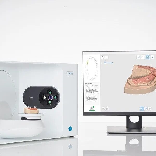
| Description | The Medit T710 desktop scanner is equipped with a fast scan engine and highly efficient software algorithm which work in tandem to produce a full-arch scan in just 8 seconds. | Image Quality
According to DEXIS, their digital xray system provides detailed image quality and includes image enhancement tools that increase diagnostic capabilities. When the 9 evaluators were asked to rate the system’s image quality and enhancement aids, 89% rated them as excellent to very good. One of the evaluators commented that the system’s “ClearVu and image magnification allow us to diagnose very effectively.”Overall Satisfaction
When asked to rate their overall satisfaction with the DEXIS Digital X-ray System, 89% of the evaluators rated it as excellent to very good. Commented one evaluator, “DEXIS has been very responsive to our needs.” | The DEXIS Digital X-ray System includes a highend CCD sensor, portable PC card and DEXusb connection options, DEXIS imaging software, a complete set of sensor holders and accessories, on-site training, a printed instruction manual, training tutorials on CD, a quick reference guide for the operatory, and 1 year of DEXcare support. | The DWX-4W goes beyond dry milling and is the epitome of wet grinding technology. It uses 4-axis simultaneous movements to wet grind precision inlays, onlays, crowns, veneers and short-span bridges from glass ceramic and composite resins. | iRay D4 has made life easy for the dental professionals around the world. Dental digital radiography is one of the current armamentarium which has polished the quality of dental treatment procedures. Portable IRAY D4 is now ready to replace the previous versions of intraoral cameras. | The DWX-51D 5-Axis Dental Milling Machine is designed for effortless production of dental prosthetics. Built for precision, error-free milling, you don’t need any machine expertise to operate it. With touch-button controls and automated milling features, the production of high-quality bridges, crowns, inlays, onlays, abutments and other restorations out of 98 mm discs and blocks has been made virtually foolproof. |
| Content | Medit T710 Blue Light Dental Scanner
Introducing the New and Improved T-Series Lab Scanners
Medit have completely overhauled the design of their T-Series lab scanners. The result is a sleek and sophisticated dental scanner which not only does its work well, but in style.The Medit T710 desktop scanner is equipped with a fast scan engine and highly efficient software algorithm which work in tandem to produce a full-arch scan in just 8 seconds. The advanced, high-speed positioning system of the new T-Series is designed for optimal performance for your laboratory, speeding up your workflow and increasing productivity.The T710, with its new four 5.0MP-resolution cameras system and blue-light scanning technology, ensures your models and impressions are fully scanned, eliminating all blind spots.Flexible multi-die scanning
2 StepsT710 Specifications
Accuracy 4μm
Full Arch Scan 8 seconds
Full Arch Impression Scan Speed 45 seconds
Colour Texture
Auto-elevation
Let the scanner decide the scanning height for your object with our auto-elevation feature.
Wider Scan Area
Scan more objects at the same time thanks to the wider scan area of our T-Series tabletop scanners!
No Blind Spots
The 4 cameras in the T710 are positioned in a way to ensure that there are no blind spots in your scan data. It only takes one scan to get the full data!
Full-size articulator scanning
To reproduce the exact occlusion orientation, nothing beats scanning the occlusion in the articulator itself. We’ve designed our T-Series to accommodate any articulator available in the market, comfortably.
Replica Denture Included
Orthodontic Scanning Included
Impression Scanning IncludedMedit T710 Blue Light Dental Scanner- ultra-fast scanning of a dental arch in only 8 seconds
- scanning speed for a complete jaw impression is only 45 seconds
- color texture available
- accuracy of 4 µm
- open System
- 5.0 megapixel camera enables high-resolution and detailed scan data
- four-camera system for a large scanning area without blind spots
- automatic height adjustment for optimal scan Position
- flexible multi-die scanning
- complete scan in the articulator possible with the 3-in-1 KAS holding device for KaVo, Artex and SAM
- the AM holding device is compatible with the MARK330 and BIOART A7+ articulators - thus the use of the exocad software is possible
- orthodontic scanning and impression scanning possible
| Dexis Intraoral Digital Dental X-Ray SensorProduct DescriptionDexis Intraoral Digital Dental X-Ray SensorSoftware Ease of Use
According to the manufacturer, the DEXIS x-ray software was designed to be highly effective throughout diagnosis, treatment planning, and treatment. It is intended to serve as an imaging hub, managing all digital images, including intra- and extraoral radiographs and photographs. The manufacturer states that the images can be quickly organized, stored, retrieved, printed, and shared with patients and colleagues. The 9 dentists who participated in this product evaluation were asked to rate the DEXIS Digital Imaging software on its ease of use with regard to taking and accessing radiographs and for diagnosis, patient education, and communication with colleagues and insurance companies. Six of the evaluators rated the system as excellent and 3 rated it as very good. One evaluator commented that “when communicating with patients, we use DEXIS images displayed on a monitor in front of the patient every day on virtually every patient.” Software Integration
The manufacturer claims that the DEXIS imaging software can be easily integrated with all major practice management systems. According to DEXIS, the DEXIS Integrator automatically synchronizes with the patient record.When the evaluators were asked to rate the software integration with practice management software and interface with hardware (eg, cameras, panoramics, and scanners), 78% rated it as excellent to very good, while 22% rated it as good. One of the evaluators said, “The bridge to our practice management systems is seamless. We use [the] DEXIS digital pan[oramic] so that we can store panoramic x-rays easily in the same location as full series. We use an intraoral camera to store preoperative and interoperative photographs for excellent documentation.” Sensor Comfort and Ease of Use
According to DEXIS, the heart of their digital system is the PerfectSize sensor. The manufacturer states that their single-size sensor has 3 advantages:
1) because the electronics are on the back of the sensor, the active imaging area is most of the total sensor size;
2) the beveled corners, slim profile, and rounded casing allow for accurate placement and patient comfort;
3) patented cord placement allows the practitioner to take vertical and horizontal bitewings and all periapicals with only 1 universally sized sensor.
The evaluators were asked to rate both the ease of use of the sensor and patient comfort. Five evaluators rated ease of use excellent and 4 rated it very good. With regard to patient comfort, 3 evaluators rated the sensor excellent and 6 rated it very good. One evaluator noted that he experienced a few issues when placing the sensor in some children and patients with very small mouths. Another evaluator, however, is very happy with the sensor, saying, “I chose DEXIS to take a full radiographic series on every patient. It was the only direct image system we tested that met that objective.” Image Quality
According to DEXIS, their digital xray system provides detailed image quality and includes image enhancement tools that increase diagnostic capabilities. When the 9 evaluators were asked to rate the system’s image quality and enhancement aids, 89% rated them as excellent to very good. One of the evaluators commented that the system’s “ClearVu and image magnification allow us to diagnose very effectively.” Overall Satisfaction
When asked to rate their overall satisfaction with the DEXIS Digital X-ray System, 89% of the evaluators rated it as excellent to very good. Commented one evaluator, “DEXIS has been very responsive to our needs.” System Includes* a highend CCD sensor
* portable PC card and DEXusb connection options
* DEXIS imaging software
* a complete set of sensor holders and accessories
* on-site training
* a printed instruction manual
* training tutorials on CD
* a quick reference guide for the operatory
* 1 year of DEXcare support. | Dexis Intraoral Digital Dental X-Ray Sensor
Software Ease of Use
According to the manufacturer, the DEXIS x-ray software was designed to be highly effective throughout diagnosis, treatment planning, and treatment. It is intended to serve as an imaging hub, managing all digital images, including intra- and extraoral radiographs and photographs. The manufacturer states that the images can be quickly organized, stored, retrieved, printed, and shared with patients and colleagues. The 9 dentists who participated in this product evaluation were asked to rate the DEXIS Digital Imaging software on its ease of use with regard to taking and accessing radiographs and for diagnosis, patient education, and communication with colleagues and insurance companies. Six of the evaluators rated the system as excellent and 3 rated it as very good. One evaluator commented that “when communicating with patients, we use DEXIS images displayed on a monitor in front of the patient every day on virtually every patient.”Software Integration
The manufacturer claims that the DEXIS imaging software can be easily integrated with all major practice management systems. According to DEXIS, the DEXIS Integrator automatically synchronizes with the patient record.
When the evaluators were asked to rate the software integration with practice management software and interface with hardware (eg, cameras, panoramics, and scanners), 78% rated it as excellent to very good, while 22% rated it as good. One of the evaluators said, “The bridge to our practice management systems is seamless. We use [the] DEXIS digital pan[oramic] so that we can store panoramic x-rays easily in the same location as full series. We use an intraoral camera to store preoperative and interoperative photographs for excellent documentation.”Sensor Comfort and Ease of Use
According to DEXIS, the heart of their digital system is the PerfectSize sensor. The manufacturer states that their single-size sensor has 3 advantages:
1) because the electronics are on the back of the sensor, the active imaging area is most of the total sensor size;
2) the beveled corners, slim profile, and rounded casing allow for accurate placement and patient comfort;
3) patented cord placement allows the practitioner to take vertical and horizontal bitewings and all periapicals with only 1 universally sized sensor. The evaluators were asked to rate both the ease of use of the sensor and patient comfort. Five evaluators rated ease of use excellent and 4 rated it very good. With regard to patient comfort, 3 evaluators rated the sensor excellent and 6 rated it very good. One evaluator noted that he experienced a few issues when placing the sensor in some children and patients with very small mouths. Another evaluator, however, is very happy with the sensor, saying, “I chose DEXIS to take a full radiographic series on every patient. It was the only direct image system we tested that met that objective.”Image Quality Dexis Intraoral Digital Dental X-Ray Sensor
According to DEXIS, their digital xray system provides detailed image quality and includes image enhancement tools that increase diagnostic capabilities. When the 9 evaluators were asked to rate the system’s image quality and enhancement aids, 89% rated them as excellent to very good. One of the evaluators commented that the system’s “ClearVu and image magnification allow us to diagnose very effectively.”Overall Satisfaction
When asked to rate their overall satisfaction with the DEXIS Digital X-ray System, 89% of the evaluators rated it as excellent to very good. Commented one evaluator, “DEXIS has been very responsive to our needs.” | Roland DWX-4W Dental Milling Unit
Reliable High PerformanceIn addition to milling on X, Y and Z axes, the DWX-4W rotates pin-type blocks 360 degrees on a fourth axis (A axis) to support undercuts. The ball screw-driven 4-axis control allows for smooth, high-precision machining. The DWX-4W’s high-performance 60,000-RPM Jaeger spindle is known for its reliability and supports precision milling of glass ceramics and composite resin. With its multi-pin clamp, the DWX-4W is a wet dental milling machine that can mill up to three different pin-type materials continuously in one operation. As a result, prosthetics for up to three different patients can be produced in a single production run, saving time, labour and costs.- Simultaneous 4-axis machining
- 4-state Automatic Tool Changer (ATC)
- 60,000 RPM Jaeger DentaDrive Spindle
- CAM software bundled
- Virtual Machine Panel (VPanel)
- Fully-integrated water and coolant filtration system
- Coolant leakage & flow monitoring sensors
- Tool life monitoring
- Mail notification system
Summary Roland DWX-4W Dental Milling Unit• Maximum Convenience
• Virtual Machine Panel
• Progress and Error Alerts for Error-Free Milling
• Coolant Compartment and Water Filtration
• Clean Cutting Area for Precision Milling
• Economical Open Architecture
• CAM Software Included
• Growth Potential
• Peace of MindThe DWX-4W’s high-performance 60 000 RPM Jaeger DentaDrive spindle supports precision milling. It’s equipped to mil pin-type blocks, including glass ceramics and composite resin – acclaimed in the industry for their superior strength and aesthetics. materials can be finished with a short finishing tome, dramatically reducing labour and production time.In addition to milling on X, Y and Z axes, the DWX-4W rotates pin-type blocks 360 degrees on a fourth axis (A axis) to support undercuts, the rigid ball screw-driven 4-axis control allows for smooth, high-precision machining. With its multi-pin clamp, it can mil up to three different pin-type materials continuously, enabling production three different patient prosthetics in a single production run. it also features an Automatic Tool Changer (ATC) that auto-changes grinding burs without interrupting the production process. | Dexcowin IRay D4 Dental Handheld X-Ray System
Dexcowin’s iRay D4 is a total dental imaging solution. The iRay D4 comes with an attachable digital sensor, a built in computer for image processing and a 4.8” digital screen so you can see your radiographs live. Diagnosis with a single machine, completely safe and mobile allows imaging on the go. The iRay D4 works with film, digital sensors and PS (photostimulable phosphor) plates. The iRay D4 is wireless, handheld, and completely mobile thus eliminating the need for designated X-ray machine rooms.Dexcowin’s iRay D4 is an all in one solution. It is an x-ray source, a computer, and comes with a high-quality sensor. The iRay D4 can be used as a simple x-ray source when used with film or a sensor attached to a separate computer.The iRay D4 is wireless, handheld, and completely mobile thus eliminating the need for designated X-ray machine rooms. The iRay D4 is perfect for the endodontic who need to surgical procedures in process or mobile dentist practicing in remote locations without computer access.Safety* Safe to hold by hand. 1/7 the radiation exposure then conventional systems.
* Rechargeable Li-poly batteries allow up to 300 images on a single charge.
* Works with any radiographic medium: film, digital sensor, PS plates.
* Operator Safe: Certified as SAFE TO USE BY HANDMobility* The iRay D4 is a complete mobile system. It has an on board computer and a 4.8” screen that allows the doctor to take analyze X-rays with a single piece of equipment.
* There is no need for additional equipment as the iRay D4 comes complete with a high definition sensor.
* The iRay D4 is the most mobile and independent solution on the market today.
* The iRay D4 comes with two rechargeable Li-poly batteries with up to 300 shots on a single charge.
* The iRay D4 also includes wireless transfer to your PC, now take and view your X-rays anywhere!Quality* The iRay D4 gives you more for less than conventional systems. The iRay D4 does not need special construction in terms of shielded rooms, chemical development and dedicating rooms for X-ray.
* The iRay D4 does not require special technical equipment such as computers and additional sensors thereby providing quality all-in-one equipment.
* It allows you to take an x-ray in real time, while you are in the room. This allows the practitioner to provide his customer face to face reassurance while the X-rays and images are taken.Still not convinced? We also offer a 5-jointed stand arm for which you can mount the iRay D systems. Comes fixated to a mobile cart so you still preserve the portable characteristics of the iRay system. With an included remote control, satisfy any operator who still feels uneasiness with being in the same room as an X-ray machine.Digital X-ray Specifications:Dexcowin IRay D4 Dental Handheld X-Ray SystemProduct Name : iRay D4
CPU : 1.6GHz
DISPLAY : 4.8”Touch Panel TFT-LCD (1024×600)
MEMORY : 16GB
EXPOSURE : 0.05 ~ 1.35, 0.01 step
BATTERY : rechargeable 4 cell high capacity battery pack
INTERFACE : USB2.0
Tube Voltage : 60kV(fixed)
Tube Current : 2mA(Fixed)
Focal Spot Size : 0.8mm
Voltage Consumption : 240W Max
Cooling Method : Oil Cooling
Total Filtering : Over 1.5mmAl (inherent filtration : 0.8mmAl, fixed type added filter : 0.7mmAl)
Target Angle : 20°
Time Setting Function : 0.05 ~ 1.35 second (0.01 step)
Main Body Size : 240(L) x 166(W) x 82(H) [mm]
Main Body Weight : 2.4kg
Battery Pack : Cell Material Quality Lithium Polymer
Charging Condition : Charging Current 1A
Temperature Range : 0~40°C
Maximum Current : 18A
Out Voltage : DC 15V
Temperature Range : -20 ~ 60°C | Roland DWX-51D Dental Milling Unit
The DWX-51D 5-Axis Dental Milling Machine is designed for effortless production of dental prosthetics. Built for precision, error-free milling, you don’t need any machine expertise to operate it. With touch-button controls and automated milling features, the production of high-quality bridges, crowns, inlays, onlays, abutments and other restorations out of 98 mm discs and blocks has been made virtually foolproof.- 5-axis simultaneous milling for complex prosthetics and full mouth restorations
- Precision milling of wax, zirconia, PEEK, gypsum and PMMA
- 10-station Automatic Tool Changer (ATC) with tool length sensor
- Built-in dust collection system for maximum extraction and easy cleaning
5-Axis Machining for Smoother Production
The DWX-51D is a smooth and steady ball screw-driven machine capable of milling complex restorations. It operates on X,Y, and Z axes – simultaneously rotating blocks and discs. Tilting on the B axis it supports deep undercuts and the complex milling of large arch restorations and other full-mouth prosthetics.Clean and Comfortable Operation
A unique inner structure with spacious airflow supports comfortable operation, while an automatic dust collection feature boasts a powerful vacuum for clean usage and calibration. An ionizer removes static, ideal for milling PMMA material.Economical Open Architecture
The open architecture of the DWX-51D allows you to work with the latest materials, CAD/CAM software and scanners, without needing to rely on a single source.VPanel for Simplified Operation
Complex controls associated with CNC milling have been replaced by a user-friendly Virtual Machine Panel (VPanel) that automatically starts the milling process, conducts machine diagnostics and operates milling calibration from your computer.New C-Clamp for Quicker Set-Up
The new C-clamp on the DWX-51D requires the adjustment of only two screws for a simplified set-up. The C-clamp (which includes a torque driver for improved clamping consistency) also provides exact alignment and uniformity, resulting in less misalignment issues.10-Station Automatic Tool Changer for Continuous Milling With a 10-station Automatic Tool Changer (ATC), DWX-51D users don’t have to stop production to replace the specific burs needed for milling different materials.User Replaceable Spindle Roland DWX-51D Dental Milling Unit
When the spindle needs replacing on the DWX-51D, there’s no need to stop production and pay for a technician to come out. Featuring a spindle that users can replace themselves, production never has to come to a complete standstill.Progress and Error Alerts for Error-Free Milling
To further automate the milling process and ensure error-free production, users receive e-mail notifications detailing any errors, or when milling is completed, while a color-coded LED light signals machine status.9 Pin-Type Material Adapter Included
A material adapter with nine pin-type placement options is included for easy and error free set-up.VITA Certified
VITA certifies the Roland DWX-51D for milling VITA ENAMIC. The DWX-51D is the only dry-milling system that, when equipped with our ZBD-100D-HY /50D-HY /30D-HY hybrid tools, is authorized to mill VITA ENAMIC and produce a full-range of dental prosthetics. Other material brands are also available for milling with the DWX-51D.Peace of Mind, Reliability and Support
The DWX-51D offers unmatched reliability and post-sales support that includes a two-year warranty. Enjoy the benefit of certified service engineers to ensure that your device is always up-and-running and a dedicated online and telephone support staff who can answer all your questions and concerns. |




Reviews
There are no reviews yet.