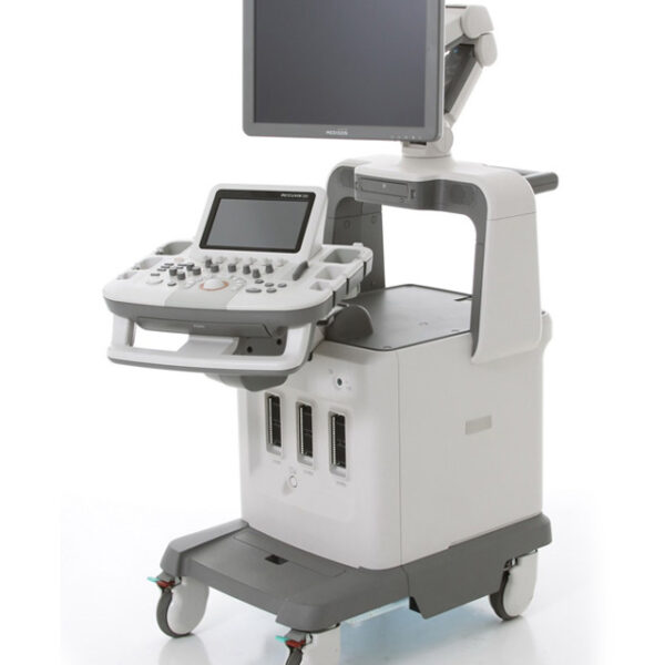
| Description | The Samsung Accuvix XG was the top of the line system from Samsung prior to the release of the newer. The XG was meant as a direct replacement to the popular Accuvix XQ as a shared service system capable of both 4D OBGYN images as well as full cardiac scans. | The GE Vivid q portable ultrasound machine is a more powerful version of the Vivid i that adds further quantitative features including ICE, Auto EF, AFI, TVI,TT, TSI, Blood Flow and Blood Flow imaging. The Vivid q is the pinnacle of GE’s portable cardiovascular ultrasounds. | GE Voluson 730 Expert is among the top-regarded ultrasounds in the industry. With up to 40 volumes per second rate of acquisition and construction of volumetric images in real time. The 730 Expert is loaded with the newest ultrasound technology including: CrossXBeam, Speckle Reduction Imaging, Tomographic Ultrasound Imaging (TUI), Spatio-Temporal Image Correlation (STIC), and much more. | The Samsung Accuvix XG was the top of the line system from Samsung prior to the release of the newer. The XG was meant as a direct replacement to the popular Accuvix XQ as a shared service system capable of both 4D OBGYN images as well as full cardiac scans. | Ultraformer III’s 7 clip-in transducer cartridges project uniform ultrasound beams directly to multiple layers underneath the skin to promote tighter collagen formation, tissue contraction and reduction in the volume of adipocytes that form bulging areas of tissue.Patients notice an immediate benefit in improved skin tone and texture, with ongoing improvements over 3 to 6 months
Traditionally difficult areas such as eye lifting are made easier with Ultraformer III’s specialised cartridges. It is possible for patients to experience up to 6mm of eyebrow lift, depending on their particular situation. | The New Terason uSmart 3200T Ultrasound System, the first in Terason’s uSmart family of proprietary products. This revolutionary new tablet, deemed “disruptive technology” by industry leaders, will change the way healthcare is delivered in both hospital and outpatient sectors of point-of-care ultrasound. |
| Content | Medison Accuvix Xg Utrasound System
SAMSUNG ACCUVIX XG ULTRASOUND MACHINE2D Image Features
• Dynamic MR / Dynamic MR Plus is designed to enrich gray-scale resolution, as it enhances detection
and contrast resolution while also decreasing speckle echoes. This is particularly useful when evaluating
superficial structures, including thyroid, vessels, pelvic and abdominal anatomy.
• SRF (Speckle Reduction Filter) enhances image quality by reducing or eliminating the appearance of
speckle echoes from ultrasound images. The degree of speckle reduction implemented is user-selectable.
• Wide Dynamic Range determines the number of gray shades utilized to map the gray-scale image.
It enables display of more details of bright areas and dark areas.
• SCI (Spatial Compounding Image) controls the ultrasound beam electronically by steering, and compounds
many scan lines.
• FSI (Full Spectrum Imaging) incorporates the penetration capabilities associated with lower frequencies,
yet maintains the fine pixel uniformity associated with higher frequencies, to deliver consistently high quality
images even in challenging diagnostic areas.3D Image Features
• HDVI gives outstanding image quality and naturally clearer contrast, with excellent tissue differentiation,
edge depiction and speckle reduction, allowing consistent diagnoses with great confidence.
• 3D XI, comprised of a suite of three innovative imaging applications – Multi-Slice View, Oblique View™ and
Volume CT – offers complete and precise control over 3D/4D volume data manipulation for maximum
diagnostic accuracy.
• 3D MXI is an innovative, cutting-edge 3D image processing technology. Comprising a comprehensive suite of
imaging tools – including Multi Volume Slice, Mirror View, Multi-OVIX, and 3D OH – 3D MXI lets you view,
examine and diagnose 3D volume data with supreme ease, speed and accuracy. • Fully adjustable system Medison Accuvix Xg Utrasound System
The control panel can be adjusted to the user’s preferred height, for a better working environment and reduced
risk of back pain.
• Wide LED touch-screen
The Accuvix XG’s new LED touch-screen makes it easy to organize and operate.
• 19-inch HD LCD Monitor and articulating monitor arm
A 19-inch LCD monitor enables images to be displayed clearly even with a larger monitor, and the articulating
monitor arm enables easy mobility for a more comfortable and convenient working environment.
• Portability
The Accuvix XG is a lightweight system with 4 swivel wheels that allow easy steering, and a locking function. • Customizable measurement menus
Customizable measurement menus allow access to frequently-used functions, and enable a quicker and
more intuitive workflow.
• User keys and user knob
Accuvix XG offers user keys and a user knob that can map frequently-used functions, enabling the function to be
activated quickly and easily.
• Customized Annotation Menu and body marker
Users can preset up to 360 words of annotations, and body markers for each application, that reduce the time
needed for each examination.
• QuickScan
QuickScan maximizes workflow efficiency by automatically optimizing key imaging parameters with just the
push of a button. • HDVI
HD Volume Imaging(HDVI) gives outstanding image quality and naturally clearer contrast, with excellent tissue
differentiation, edge depiction and speckle reduction, allowing consistent diagnoses with great confidence.
It shows clearer images of subtle lesions, using multi slice image as well as C-plane.
• ElastoScan
ElastoScan™ translates stiff & soft tissue information into gray scale image. This technology defines the alteration
of the stiffness in tissue state more accurately, making it easier for the user to utilize the information in the image. | GE Vivid Q Ultrasound Machine
The powerful GE Vivid Q ultrasound machine is the ultimate lightweight, compact high-performance cardiovascular system for clinicians needing a machine that combines user efficiency and reliable performance. This unique system performs consistently thanks to its TruScan architecture and can support up to 17 probes. The GE Vivid Q is recognized for its premium image quality, advanced image management, and high-performance quantification tools.This advanced GE ultrasound delivers improved patient care and enhanced user productivity with the new M4s-Matrix array technology transducer, which provides greatly enhanced image quality in electronic beamforming, raw data image acquisition and archiving, optimal onboard and post processing technology.This ultrasound system provides high clinical values for quantification and review of stored images, making it one of today’s most sought-after models. Some of its powerful features include compatibility with EchoPac clinical workstation and DICOM networking, speckle tracking strain, integrated stress echo, quantification tools, and reporting capabilities. Also of note is one of the Vivid Q’s best new features, which is its TEE transducer capability.A one-touch button optimization provides premium image quality for workflow efficiency, and maintains AFI- automatic function imaging with complete wallFeatures- Advanced QScan Imaging (incl TSI)
- AdvStress
- ATO
- Auto EF
- Automated Function Imaging
- Blood Flow Imaging
- CardiacApp
- Cardio Lab
- Color Doppler
- CW Doppler
- DICOM Networking
- DICOM: Print
- DICOM: Worklist
- DVD/RW
- ECG
- ECG Cable
- EchoDicom
- EchoPAC
- Evue
- ICE Imaging
- Intima Media Thickness (IMT)
- LVO Contrast
- M Mode
- M4S
- MPEGvue
- PW Doppler
- Small Depth
- TEE
- THI
- Tissue Harmonics Imaging (THI/NTHI)
- Tissue Velocity Imagine & Tissue Tracking
- TSI
- VascAbdContrast
- VascularApp
- VirtualPrinter
- Wide Aperture
GE Vivid Q FeaturesThe GE Vivid Qis a Portable Laptop-style Ultrasound that can be used to help diagnose cardiovascular anatomy. Features and tools based on the previous Vivid 7 and the Vivid E9 have been added to the new lightweight Vivid Q, mainly the M4S-RS transducer that allows for 3D image focusing and enables a higher level of image quality. The Vivid Q ultrasound is versatile and can additionally be used for Cardiac, vascular, Abdominal, Pediatric, Obstetrics, and Emergency medicine. The Vivid Q Ultrasound comes with a 15-inch color LCD monitor that can display a variety of imaging modes including; ICE, Auto EF, AFI, TVI, TT, TSI, and Blood Flow. Additional the Ultrasound system is comparable with 17 different transducers and probes to fit every imaging need. - Auto EF - Automated ejection fraction measurement program designed specifically for the GE Vivid q.
- Tissue Synchronization Imaging (TSI).
- Intracardiac Echo (ICE).
- 16 different compatible probes.
- Coded Octave imaging - enhances image color flow and Doppler imaging.
| GE Voluson 730 Expert Ultrasound MachineGE Voluson 730 Expert, 3D/4D Realtime Ultrasound System Color Doppler, PW Doppler, Tissue Harmonics, STIC, VOCAL DICOM, M-mode, Angio, ATO, TUI, SonoView II, Crossbeam (CRI) MO Drive, Cineloop, High Resoluotion Interlaced Monitor Probe Options: RAB4-8 4D Convex RIC5-9 4D Transvaginal AB2-7 2D Convex IC5-9 2D Transvaginal SP6-12 2D Linear PA2-5 Cardiac Sector
Two Probes included:- RAB4-8L – Live 4D Abdominal Transducer
- RIC5-9 – Live 4D Transvaginal Transducer
Options:
About the Voluson 730:GE Voluson 730 Expert Ultrasound MachineWith GE Voluson 730, you can now acquire and construct volumetric images in real time – up to 40 volumes per second. The system allows you to explore images in any plane to reveal the smallest details with stunning clarity and apply sophisticated analytical tools to answer virtually any of your clinical questions. Volume Ultrasound not only improves your 3D/4D capabilities, it enhances your 2D imaging as well, for even greater diagnostic confidence in your obstetrical, gynecologic, breast and general imaging studies.The used GE Voluson 730 Expert was among the top-rated and biggest breakthrough ultrasounds in the industry. With up to 40 volumes per second rate of acquisition and construction of volumetric images in real time. The 730 Expert is loaded with the newest ultrasound technology including: CrossXBeam, Speckle Reduction Imaging, Tomographic Ultrasound Imaging (TUI), Spatio-Temporal Image Correlation (STIC), and much more.The Voluson 730 Expert is the more advanced version of the Voluson 730 Pro and was replaced by the GE Voluson e8. Compared to the 730 Pro, the Voluson 730 Expert features a touchscreen interface, higher frame rates in 2D and 4D, and more advanced imaging options such as STIC and TUI.Providian Medical Voluson 730 Expert Review:The Voluson 730 is a workhorse of a 4D ultrasound machine. It remains a popular system in many OB/GYN/Women’s Health offices, as well as 4D “Babyface” studios around the world. It is a durable machine with very good image quality for all women’s health needs. | Medison Accuvix XG Ultrasound System
Samsung Accuvix XG Ultrasound Machine2D Image Features
• Dynamic MR™ / Dynamic MR Plus is designed to enrich gray-scale resolution, as it enhances detection
and contrast resolution while also decreasing speckle echoes. This is particularly useful when evaluating
superficial structures, including thyroid, vessels, pelvic and abdominal anatomy.
• SRF (Speckle Reduction Filter™) enhances image quality by reducing or eliminating the appearance of
speckle echoes from ultrasound images. The degree of speckle reduction implemented is user-selectable.
• Wide Dynamic Range determines the number of gray shades utilized to map the gray-scale image.
It enables display of more details of bright areas and dark areas.
• SCI (Spatial Compounding Image) controls the ultrasound beam electronically by steering, and compounds
many scan lines.
• FSI (Full Spectrum Imaging) incorporates the penetration capabilities associated with lower frequencies,
yet maintains the fine pixel uniformity associated with higher frequencies, to deliver consistently high quality
images even in challenging diagnostic areas.3D Image Features Medison Accuvix XG Ultrasound System
• HDVI gives outstanding image quality and naturally clearer contrast, with excellent tissue differentiation,
edge depiction and speckle reduction, allowing consistent diagnoses with great confidence.
• 3D XI™, comprised of a suite of three innovative imaging applications – Multi-Slice View, Oblique View™ and
Volume CT – offers complete and precise control over 3D/4D volume data manipulation for maximum
diagnostic accuracy.
• 3D MXI is an innovative, cutting-edge 3D image processing technology. Comprising a comprehensive suite of
imaging tools – including Multi Volume Slice, Mirror View, Multi-OVIX, and 3D OH – 3D MXI lets you view,
examine and diagnose 3D volume data with supreme ease, speed and accuracy. • Fully adjustable system
The control panel can be adjusted to the user’s preferred height, for a better working environment and reduced
risk of back pain.
• Wide LED touch-screen
The Accuvix XG’s new LED touch-screen makes it easy to organize and operate.
• 19-inch HD LCD Monitor and articulating monitor arm
A 19-inch LCD monitor enables images to be displayed clearly even with a larger monitor, and the articulating
monitor arm enables easy mobility for a more comfortable and convenient working environment.
• Portability
The Accuvix XG is a lightweight system with 4 swivel wheels that allow easy steering, and a locking function. • Customizable measurement menus
Customizable measurement menus allow access to frequently-used functions, and enable a quicker and
more intuitive workflow.
• User keys and user knob
Accuvix XG offers user keys and a user knob that can map frequently-used functions, enabling the function to be
activated quickly and easily.
• Customized Annotation Menu and body marker
Users can preset up to 360 words of annotations, and body markers for each application, that reduce the time
needed for each examination.
• QuickScan
QuickScan maximizes workflow efficiency by automatically optimizing key imaging parameters with just the
push of a button. • HDVI
HD Volume Imaging(HDVI) gives outstanding image quality and naturally clearer contrast, with excellent tissue
differentiation, edge depiction and speckle reduction, allowing consistent diagnoses with great confidence.
It shows clearer images of subtle lesions, using multi slice image as well as C-plane.
• ElastoScan
ElastoScan™ translates stiff & soft tissue information into gray scale image. This technology defines the alteration
of the stiffness in tissue state more accurately, making it easier for the user to utilize the information in the image. | CLASSYS ULTRAFORMER III UTR3
You’ve heard about it, now see and feel the difference for yourself.Ultraformer III is the leading technology for facial lifting, tightening and contouring. Now with 7 interchangeable cartridges that offer your patients another dimension in non-surgical face and body treatments.Ultraformer III is widely used with over 10 million treatments performed worldwide. It has become the treatment of choice for patients who are not yet indicated or not willing to undergo surgery. This fills a huge gap in the market, and captures an audience of people from 30 years of age all the way to 80.ULTRAFORMER III FEATURESLess discomfort associated with treatments
Patented transducer design for more efficient energy delivery
Dual hand pieces with automatic recognition for ease of use
Faster shot and treatment speed to save time
Easy, user-friendly design and interface
Shot count information and voice assistance
Backed by clinical papersThe CLASSYS ULTRAFORMER III UTR3 advantage with slim transducer designimproves operator visibility
supports very narrow focus and precise treatment
fits facial contoursFast elliptic transducersmore efficient treatment timesUltraformer III’s 7 clip-in transducer cartridges project uniform ultrasound beams directly to multiple layers underneath the skin to promote tighter collagen formation, tissue contraction and reduction in the volume of adipocytes that form bulging areas of tissue.Patients notice an immediate benefit in improved skin tone and texture, with ongoing improvements over 3 to 6 months
Traditionally difficult areas such as eye lifting are made easier with Ultraformer III’s specialised cartridges. It is possible for patients to experience up to 6mm of eyebrow lift, depending on their particular situation.Ultraformer III’s 7 clip-in transducer cartridges project uniform ultrasound beams directly to multiple layers underneath the skin to promote tighter collagen formation, tissue contraction and reduction in the volume of adipocytes that form bulging areas of tissue.Patients notice an immediate benefit in improved skin tone and texture, with ongoing improvements over 3 to 6 months
Traditionally difficult areas such as eye lifting are made easier with Ultraformer III’s specialised cartridges. It is possible for patients to experience up to 6mm of eyebrow lift, depending on their particular situation. | Terason uSmart 3200t Ultrasound
The new light-weight, power-packed uSmart 3200T Ultrasound System is a fusion of cutting-edge technology with an intuitive interface that defines simplicity. The newest member of the Terason family weighs less than 5 lbs., provides razor-sharp image quality, and is fully equipped with all the features and functionality users have come to expect from Terason products. Developed by clinicians for clinicians, the uSmart 3200T provides the best tools for busy practices, including a 128GB Solid State hard drive, Smart Gestures, an Adaptive Touch Screen, uConnect remote capabilities, and a fast boot-up time.The Terason uSmart 3200T Ultrasound is truly a unique and revolutionary product in the world of portable ultrasound. In our fast-paced technology environment, healthcare providers require products that can keep up with the ever-changing demands of their hospital or practice. We have developed the first tablet-based system where high technology meets superior performanceThe Terason uSmart 3200T is a tablet-style portable color Doppler ultrasound machine designed for the point-of-care bedside ultrasound market.This handheld ultrasound weighs less than 5lbs and uses gestures and touch operation much like a tablet or touchscreen computer. With Terason’s well-known unique beamforming microprocessor technology, the Terason uSmart 3200T Ultrasound provides excellent image quality with an efficient workflow and easy use.Gold Standard, Crystal Clear Images you have to see to believe! As an industry pioneer, Terason developed and is the first to patent color portable ultrasound. Since then, our razor-sharp image quality has earned a reputation for excellence.
The uSmart 3200T captures every detail, subtlety, and nuance that others overlook. The AHVA LCD provides consistent image quality from multiple viewing angles.Grab-And-Go Portability
As part of one of the most comprehensive product families in the industry, the uSmart 3200T weighs in at just under 5 pounds. The touchscreen interface, small footprint an space-saving power dock can go anywhere you need quickly and easily. It’s ready when you are! The Best Tools For Your Practice- Small Gestures
- Adaptive touch screen
- uConnect remote capabilities
- Multi-Function docking station
- Solid State Hard Drive
- Application-Specific presets
- Fast boot-up time
Enhanced Needle Visualization (ENV)
With ENV- AS a leader in ultrasound, Terason is pleased to introduce ENV. This feature provides needle visualization unlike any other. - Frame rate stability
- One-button operation
- Works with all needle gauges
- 15L transducer solution
- Easy to use
Terason uSmart 3200T Ultrasound Specifications- Dimensions: 22.4 cm x 3. 2 cm x 3.21 cm
(8.82″ x 1.26″ x 12.64″)
- 12.5″ x 1.25″ x 8.75″ High-Res AHVA LCD – Touch Screen
- 128 GB Hard Drive
- Input / Output: 3 USB-A, Ethernet & DVI
- Weight: 4.5 pounds
- Export: to USB, Network Folder
- Operates on AC or internal battery power
Imaging Modes:- B- mode
- M-mode
- Color Doppler
- Color Power Doppler
- Directional Color Power Doppler
- PW Doppler
|




Reviews
There are no reviews yet.