| Description | The image can then be displayed on a monitor, helping you to educate your patients The HRK can provide peripheral keratometry measurement data which is very useful when fitting contact lenses. The eye can be displayed in full colour mode on a tilting touch screen which enables you to see the patient’s contact lens fitting status, previously only possible using a slit lamp. As the HRK has the world’s first contact lens fitting function in an auto keratometer, you can now see the flourescein liquid with blue illumination along with automatic calculation and recommendation for contact lens fitting. | Ezer ERK-7800 Auto Refractor/KeratometerConfidently measure the human eye with a sophisticated cutting edge technology designed to enhance your ability to diagnose ocular errors faster, easier, and accurately. Ezers ERK-7800 is brilliantly designed technology measures the eye with ultra precision and speed to provide reliable data as the starting point of your refraction and contact lens fitting. It’s compact and modern design enables seamless operation and an enhanced patient comfort, making it the ideal choice in for any ophthalmic practice. Backed by an industry leading 2-years warranty! | Combination of auto and manual analysesAll three manual analysis methods can be performed on the same image and also on auto-analyzed images. The versatility of combining automated and manual analyses allows analysis of the range of pathology in a comprehensive practice.
The CEM-530 with new advanced software and enhanced image capture system allows rapid image acquisition. | The Cirrus HD-OCT is a non-contact, high resolution tomographic and biomicroscopic imaging device. It is indicated for in-vivo viewing, axial cross-sectional, and three-dimensional imaging and measurement of anterior and posterior ocular structures, including cornea, retina, retinal nerve fiber layer, ganglion cell plus inner plexiform layer, macula, and optic nerve head. The Cirrus normative databases are quantitative tools for the comparison of retinal nerve fiber layer thickness, macular thickness, ganglion cell plus inner plexiform layer thickness, and optic nerve head measurements to a database of normal subjects. The Cirrus HD-OCT is intended for use as a diagnostic device to aid in the detection and management of ocular diseases including, but not limited to, macular holes, cystoid macular edema, diabetic retinopathy, age-related macular degeneration, and glaucoma. | Confidently measure the human eye with a sophisticated cutting edge technology designed to enhance your ability to diagnose ocular errors faster, easier, and accurately. Ezers ERK-7800 is brilliantly designed technology measures the eye with ultra precision and speed to provide reliable data as the starting point of your refraction and contact lens fitting. It’s compact and modern design enables seamless operation and an enhanced patient comfort, making it the ideal choice in for any ophthalmic practice. Backed by an industry leading 2-years warranty! | Experience 6 analyses in 1 device: refraction, keratometry, topography, tonometry, pachymetry and dry eye analysis. |
| Content | Huvitz HRK-8000A Auto Refractor KeratometerHuvitz Auto Ref – Keratometer HRK-8000A- Touch and tilting 7″ LCD Screen that allows for free and smooth tilting for viewing at any angle.
- Optimized Optical System
- Micro Lens Array
- Aberration Measurement which also can be displayed in a graphical Zernike Refraction Map
- PSF & Image Simulation
- Colour View Mode
- Peripheral Keratometry Measurement
- Ultra High Precision KER Data
- Contact Lens Fitting Assistance Guide
- Contact Lens Prescription Guide
- Auto Tracking
- Animated Guide
- Faster Measurement Speed
- Quiet and speedy auto cutting printer
- More data on aberration measurement
With its extreme precision and accuracy, the HRK8000A uses the most advanced wavefront technology to measure the light reflected from the retina and helps you to diagnose any refractive error. With a micro lens array developed by Huvitz, the HRK creates a number of focal points, the pattern of which can give a valuable insight into the patients ocular system. The HRK not only displays the conventional data such as spherical, cylinder and axis, the high order aberration data is displayed in a graphical Zernike refractive map, giving you a deeper understanding of your patient’s eyes.Optimized Optical System Huvitz HRK-8000A Auto Refractor Keratometer
A wavefront is a physical representation of the optical quality of a light beam. Using various methods, aberrometers map how light rays travel through the eye. The HR-8000A is engineered according to the Hartmann-Shack wavefront measurement technique, which is widely preferred for its speed and repeatability.Touch & Tilting Color Display Screen
A bright, high contrast 7″ VGA color TFT LCD screen provides high resolution video images. A smooth, free tilting function also offers you a comfortable and clear view at any angle.The image can then be displayed on a monitor, helping you to educate your patients The HRK can provide peripheral keratometry measurement data which is very useful when fitting contact lenses. The eye can be displayed in full colour mode on a tilting touch screen which enables you to see the patient’s contact lens fitting status, previously only possible using a slit lamp. As the HRK has the world’s first contact lens fitting function in an auto keratometer, you can now see the flourescein liquid with blue illumination along with automatic calculation and recommendation for contact lens fitting. | Ezer ERK-7800 Autorefractor KeratometerFeatures of the ERK-7800 Auto Refractor/Keratometer
 Auto Refractor/Keratometer
Enhanced Retro-illumination Imaging
Accurate Measurement of Iris & Pupils Diameters
Precise Temperature Sensor Compensation
Enhanced Measuring Speed
Comfortable Motorized Chin Rest
Automatic High Speed Cutting Printer
Simple One Touch Lock Knob
20 Touch & Tilt Color Monitor Display
Sophisticated TFT LCD Graphic User Interface
Easy Network Capability
Backed by an Industry Leading 2-years Warranty!
Auto Refractor/Keratometer
The ERK-7800 instantly reports the patient’s assessed eye power by combining the Auto Refractor/Keratometer functions into this one sharp instrument, making measurement a snap. This determines the refractive and keratometric properties of the human eye to assist eye care professionals in the process of prescribing corrective eyeglasses and contact lenses. Keratometry and Refractometry (RK Mode) Enhanced Retro-illumination Imaging Ezer ERK-7800 Autorefractor Keratometer
With the Enhanced Retro-illumination Imaging capability in the ERK-7800, you can treat and see abnormal crystalline lenses, cataracts, and scratches of corneas helping you to determine how healthy the patients eyes are with better clarity. This allows for observation of the eye condition of the cataract or contact lens surface. The system saves up to 2 images of each eye. Accurate Measurement of Iris & Pupils Diameters
The corneal diameter and base curve of contact lenses can be measured with this one instrument. Therefore, measurements of eye and prescriptions for glasses and contact lenses can be made more efficiently. Measuring Cornea Size (SIZE MODE) Wide Dioptric Measurement Range
Because the ERK-7800 covers a wide measurement range from -25D to +22D, even an examinee with strong myopia can be measured.
Enhanced Measuring Speed
Specially designed image chips have succeeded to make REF measuring speed faster reducing the overall examination time and increases throughput for complete examinations. Comfortable Motorized Chin Rest
Simply by pressing up/down buttons, user can set the height of measuring point comfortably. With the cutting edge auto sensor mechanism, you can track down, measure and focus the eye with out ever making a mistake. Automatic Printer Paper Cut
The printer is designed to execute, print, & cut data automatically at high speed for immediate results. Simple One Touch Lock Knob
With just the touch of a single button, the main body can easily be locked to its base. 20 Degrees Tilted LCD Monitor
The LCD Monitor tilts up to 20 degrees; a design made for comfort making. Excellent ergonomics makes it possible to operate the ERK-7800 while standing or sitting, for added convenience. Sophisticated TFT LCD Graphic User Interface
Sharp images with easy to understand icons make this 6.4-inch color TFT LCD interface user-friendly and enjoyable to work with. Auto Refractor/Keratometer
Enhanced Retro-illumination Imaging
Accurate Measurement of Iris & Pupils Diameters
Precise Temperature Sensor Compensation
Enhanced Measuring Speed
Comfortable Motorized Chin Rest
Automatic High Speed Cutting Printer
Simple One Touch Lock Knob
20 Touch & Tilt Color Monitor Display
Sophisticated TFT LCD Graphic User Interface
Easy Network Capability
Backed by an Industry Leading 2-years Warranty!
Auto Refractor/Keratometer
The ERK-7800 instantly reports the patient’s assessed eye power by combining the Auto Refractor/Keratometer functions into this one sharp instrument, making measurement a snap. This determines the refractive and keratometric properties of the human eye to assist eye care professionals in the process of prescribing corrective eyeglasses and contact lenses. Keratometry and Refractometry (RK Mode) Enhanced Retro-illumination Imaging Ezer ERK-7800 Autorefractor Keratometer
With the Enhanced Retro-illumination Imaging capability in the ERK-7800, you can treat and see abnormal crystalline lenses, cataracts, and scratches of corneas helping you to determine how healthy the patients eyes are with better clarity. This allows for observation of the eye condition of the cataract or contact lens surface. The system saves up to 2 images of each eye. Accurate Measurement of Iris & Pupils Diameters
The corneal diameter and base curve of contact lenses can be measured with this one instrument. Therefore, measurements of eye and prescriptions for glasses and contact lenses can be made more efficiently. Measuring Cornea Size (SIZE MODE) Wide Dioptric Measurement Range
Because the ERK-7800 covers a wide measurement range from -25D to +22D, even an examinee with strong myopia can be measured.
Enhanced Measuring Speed
Specially designed image chips have succeeded to make REF measuring speed faster reducing the overall examination time and increases throughput for complete examinations. Comfortable Motorized Chin Rest
Simply by pressing up/down buttons, user can set the height of measuring point comfortably. With the cutting edge auto sensor mechanism, you can track down, measure and focus the eye with out ever making a mistake. Automatic Printer Paper Cut
The printer is designed to execute, print, & cut data automatically at high speed for immediate results. Simple One Touch Lock Knob
With just the touch of a single button, the main body can easily be locked to its base. 20 Degrees Tilted LCD Monitor
The LCD Monitor tilts up to 20 degrees; a design made for comfort making. Excellent ergonomics makes it possible to operate the ERK-7800 while standing or sitting, for added convenience. Sophisticated TFT LCD Graphic User Interface
Sharp images with easy to understand icons make this 6.4-inch color TFT LCD interface user-friendly and enjoyable to work with. | Nidek CEM-530 Specular Microscope
The analysis results with graphic and color-coded cell images helps the clinician to rapidly and effectively evaluate the endothelial cell layer.In addition to conventional central and peripheral specular microscopy, the CEM-530 includes a unique function to capture paracentral images.- Paracentral specular microscopy
- Faster measurements and two-second auto analysis
- Comprehensive analysis
- Advanced manual analysis functions
- Easy operation
- Additional features with CEM Viewer for NAVIS-EX
Combination of auto and manual analysesAll three manual analysis methods can be performed on the same image and also on auto-analyzed images. The versatility of combining automated and manual analyses allows analysis of the range of pathology in a comprehensive practice. Features- Multi area specular microscopy
- Enhanced usability and quick analysis
- Advanced manual analysis functions
- Combination of auto and manual analyses
- Additional features with CEM Viewer for NAVIS-EX
Multi area specular microscopyIn addition to conventional central and peripheral specular microscopy, the CEM-530 includes a NIDEK original function that captures paracentral images. The combination of central, paracentral, and peripheral imaging provides a broader, overall view that can be used for detailed morphological and quantitative evaluation of the endothelial layer and individual cells.
Enhanced usability and quick analysis
The 3D auto tracking and auto shot functions result in a user friendly and patient friendly experience.
Data analysis within 2 seconds allows efficient patient flow.
  Detail analysis
Advanced manual analysis functions
Center point
Select the approximate center of a cell. The cells are detected based on the surrounding points. This method is effective for areas where groups of cells are clumped together.
Corner point
Trace the outlines of the cells to be analyzed by selecting the corners of each cell. This method is suitable for detailed identification of the size and dimension of isolated cells.
Pattern select
Select a hexagonal reference pattern that is similar to the cell size and drag it onto the cell to be analyzed. This method is effective for rough identification of the size and dimension of the cells.
Combination of auto and manual analyses All three manual analysis methods can be performed on the same image and on auto-analyzed images. The versatility of combining automated and manual analysis on the same image allows for better clinical interpretation of the diverse range of pathology in a comprehensive practice.
Additional features with CEM Viewer for NAVIS-EX* (optional)
CEM Viewer is software used for viewing and working with CEM-530 data via NAVIS-EX. This function enhances the capability of the CEM-530 with additional features and increases the efficiency of any clinic.
*NAVIS-EX is optional software and is required for use of the CEM Viewer.
Data management and endothelial cell count
Unlimited NAVIS-EX database is available for review on the CEM Viewer. The basic functions of the CEM-530 such as endothelial cell count are available on the CEM Viewer.
Progression follow-up and comparison
Multiple examination data sets are displayed in chronological order for follow-up. Additionally, two data sets are displayed for comparison. Endothelial changes can be monitored over time with this function.
Paracentral display with peripheralThe images and analyses of the paracentral and peripheral areas are displayed providing a comprehensive image of endothelial cells. | Zeiss Cirrus HD-OCT 4000 OCT Spectral Domain OCT HD
Accessories- The ZEISS Certified Factory Direct Cirrus 4000 HD-OCT also includes the full Accessory Kit
- External Fixation Light
- Test Eye
- Mini Keyboard
- Optical Mouse
- Dust Cover
- Metric 5A/250v Fuse
- 14 Ft. CAT 5e Network Cable
- USB Cable
- IEC 320 to NEMA 12 inch Power Cable
- IEC 320 39in. Power Cable
- Install Disc including Roxio Basic
- Symbol Sheet
- OCT Primer Guide
- Quick Start Guide
HD-OCT Imaging- Methodology: Spectral Domain OCT
- Optical Source: superluminescent diode (SLD), 840 nm
- Optical Power: < 725 μW at the cornea
- Scan Speed: 27,000 A-scans per second
- A-Scan depth: 2.0 mm (in tissue), 1024 points
- Axial resolution: 5 μm (in tissue)
- Transverse resolution: 15 μm (in tissue)
Fundus Imaging- Methodology: Line scanning ophthalmoscope
- Live Fundus Image: During alignment and during OCT scan
- Optical Source: Superluminescent diode (SLD), 750 nm
- Optical Power: < 1.5 mW at the cornea
- Field of View: 36 degrees W x 30 degrees H
- Frame rate: >20 Hz
- Transverse resolution: 25 μm (in tissue)
Iris Imaging- Methodology: CCD Camera
- Resolution: 1280 x 1024
- Live iris image: During alignment
Electrical, Physical and Environmental- Weight: 38kg (83 lbs)
- Dimensions: 65L x 44W x 53H (cm)
- Fixation: Internal, external
- Internal fixation focus adjustment: -20D to +20D (diopters)
- Input devices: Keyboard, mouse
- Electrical rating (115V) Single Phase, 100/120V~ systems:50-60Hz, 5A
- Fuse rating (115V) T 5A 250V
- Electrical rating (230V) Single Phase, 220/240V~ systems:50-60 Hz, 2.5A
- Fuse rating (230V) Fuse rating: T 5A 250V
- Convenience Receptacle output ratings 115V~, 0.5 A Max, 50-60 Hz
- Temperature (transport and storage) -40º to +70º C
- Relative humidity (transport and storage) 10% to 100%, including condensation
- Atmospheric pressure (transport and storage): 500 hPa to 1060 hPa
- Temperature (operation) +10º to +35º C
- Relative humidity (operation) 30% to 75%, excluding condensation
- Atmospheric Pressure (operation) 700 hPa to 1060 hPa
Computer Zeiss Cirrus HD-OCT 4000 OCT Spectral Domain OCT HD- • High performance multi-core processor
- • Internal storage: > 80,000 scans
- • CD-RW, DVD-ROM drive
- • Integrated 15″ color flat panel display.
The Cirrus HD-OCT is a non-contact, high resolution tomographic and biomicroscopic imaging device. It is indicated for in-vivo viewing, axial cross-sectional, and three-dimensional imaging and measurement of anterior and posterior ocular structures, including cornea, retina, retinal nerve fiber layer, ganglion cell plus inner plexiform layer, macula, and optic nerve head. The Cirrus normative databases are quantitative tools for the comparison of retinal nerve fiber layer thickness, macular thickness, ganglion cell plus inner plexiform layer thickness, and optic nerve head measurements to a database of normal subjects. The Cirrus HD-OCT is intended for use as a diagnostic device to aid in the detection and management of ocular diseases including, but not limited to, macular holes, cystoid macular edema, diabetic retinopathy, age-related macular degeneration, and glaucoma. | Ezer ERK-9000 Ezer Autorefractor KeratometerThe ERK-9000 offers your staff the opportunity to gather a wealth of precise information about the patient in one fast and easy test, making the remainder of your exam swift, simple and exact. In addition to the wealth of functions you’d expect from a top-of-the-line autorefractor/keratometer, the ERK-9000 features auto-shoot programming, an illumination mode, IOL mode and so much more.
Features:– Auto Refractor/Keratometer
– Auto-Shoot Programming
– Simple and Convenient User Environment
– IOL Mode
– Illumination Mode
– Easy Network Capability
– Wide Dioptric Measurement Range
– Accurate Iris and Pupil Measurement
– Ergonomic Joystick
– Motorized Chin Rest
– High-Speed Printer with Auto Paper Cut
– 2-Year WarrantyIncludes:Ezer ERK-9000 Ezer Autorefractor Keratometer– Power Supply Cable
– Model Eye ( S-5.0D or 5.12D or 5.25D, VD=12mm, R–7.95 )
– 2 rolls Printing Paper( Thermal Paper, Size : 57 X 50 mm )
– Dust cover ( Vinyl Cover: Use for long-term storage to prevent dust pollution )
– Operation ManualThe ERK-9000 combines all of the necessary visual pre-testing functions in one smartly designed instrument. Operators can perform refractometry and keratometry alone or simultaneously. The ERK-9000 is also quick. In fact, not even the unwanted effects of patient accommodation will stand in your way thanks to the unit’s valuable built-in auto-fogging mechanism.Auto-Shoot Programming
Users can program the ERK-9000 to shoot at their preferred mode. One click on the joystick button can take a single measurement, three continuous measurements or five continuous measurements.Simple and Convenient User Environment
The ERK-9000 features a user-friendly dashboard view. Sharp images with easy-to-understand icons make this 7.0-inch color TFT color touch screen LCD interface enjoyable to work with.
IOLMode
Patients presenting with cataract or IOL won’t present a challenge with the ERK-7800. In fact, now you can gather a wealth of information about natural lens opacity as well as take measurements on patients who have intraocular lens implants. | Rodenstock Alino Multifunction Refractometer
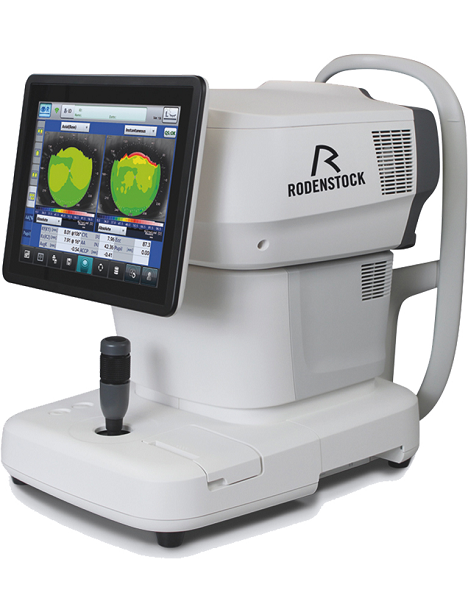
Tune-up your businessExperience 6 analyses in 1 device: Rodenstock Alino Multifunction Refractometer
Are you ready for something that is all-in-one, timesaving, space-saving and suits your business perfectly?Have a look at how powerful ALINO can be.6 in 1 solution01 RefractionHaving a good starting value for subjective refraction is essential. The QUICK REF MODE supports this even for uncooperative patients.02 KeratometryKAI (Kerato-asymmetry index) & KRI (Kerato-regularity index) give the first hint of possible irregularities on the cornea. This supports evaluation of the best vision correction or best-fitting contact lens.03 TonometryThe very gentle air puff created by a new generation of air-flow technology means that patients more readily accept the need for regular check-ups of the IOP measurement.04 PachymetryCorrecting the IOP by measuring the central corneal thickness is essential in interpreting the intraocular pressure.05 TopographyVarious topographic maps support you in carrying out vision screenings, contact lens fittings and in patient education.06 Dry EyeObserve the customers’ eye health and evaluate the long-term tolerability of contact lenses.Take advantage of the combination of tests:Additional servicesTonometry + PachymetryContact lens fitting andtolerance controlKeratometry + Topography + Dry EyeEye healthcareTopography + Tonometry + Pachymetry + Dry EyeSpecifications Rodenstock Alino Multifunction Refractometer
REFRACTIVE POWER MEASUREMENTSpherical refractive power (S) -30.00 D to + 25.00 D (at VD = 12.0 mm)Cylindrical power (C) 0.00 D to ± 12.50 D (at VD = 12.0 mm)Astigmatic axis (A) 0° to 180°KERATOMETRY MEASUREMENTCorneal curvature radius 5.00 mm to 13.00 mmCorneal astigmatic axis 0° to 180°INTRAOCULAR PRESSURE MEASUREMENTMeasurement range 1 mmHg to 60 mmHG (1 hPa to 80 hPa)PACHYMETRY MEASUREMENTMeasurement range 300 μm to 800 μmTOPOGRAPHY MEASUREMENTCorneal curvature radius 5.50 mm to 10.00 mmCorneal astigmatic axis 0° to 180°AUXILIARY FUNCTIONInterpupillary distance Measurement range: 20 mm to 85 mmCorneal diameter and pupil diameter Measurement range: 1.00 mm to 14.00 mmDry eye observation Blinking Analysis, Tear Meniscus height, Hyperemia, Meibomian glandsDATA MANAGEMENTInternal Database Integrated SD CardPrinter Integrated thermal printerData Output Type 3x USB 2.0, 1x LAN, 1x SD Card-slot, 1x WLANExport Format CSV, XML, JPG, PDFDIMENSIONS & ELECTRIC REQUIREMENTSDimensions WDH 312 × 491 × 450 mmWeight Approx. 23 kgVoltage 100 VAC to 240 VACFrequency 50/60 HzPower consumption 110 VA |




Reviews
There are no reviews yet.