| Description | Comfortable and Durable.Due to technological advances with the optical plate and scintillator, all size sensors have increased "sensitivity," which allows practitioners to decrease the radiation dose €” up to 10% decrease for sizes 1 & 2 and 40% decrease for size 0 (depending on type and age of x-ray generator). In addition, the sensor's rounded corners and ergonomic design helps make positioning easier than ever. | The DEXIS Digital X-ray System includes a highend CCD sensor, portable PC card and DEXusb connection options, DEXIS imaging software, a complete set of sensor holders and accessories, on-site training, a printed instruction manual, training tutorials on CD, a quick reference guide for the operatory, and 1 year of DEXcare support. | As one of the most user-friendly Cone Beam Computed Tomography (CBCT) systems on the market, the 9000 3D Extraoral Imaging System offers several versatile solutions for your extraoral imaging needs. Combining focused-field 3D technology with dedicated panoramic imaging, this affordable 2-in-1 unit delivers the best of both worlds, offering the highest resolution and the lowest radiation dose. | Schick Technologies has brought digital panoramic imaging to a whole new level of affordability and simplicity. | The 3Shape D710 is optimized for scanning both the impression and gypsum models and enables high throughput for both advanced and standard indications. These innovative scanners are so easy to use that staff training is barely required. Item fixation is fast, and the scan starts with a click on the program. | SuniRay 2 achieves maximal image quality with superior diagnostic capabilities while maintaining the lowest radiation levels among all digital sensors. The comprehensive and feature-rich Prof. Suni Advanced Imaging Software supplements the SuniRay 2’s high-quality imaging with a wide array of user-friendly customization and image-enhancement tools. With the SuniRay 2, Suni has created a complete digital radiography system that seamlessly integrates with your entire practice. |
| Content | Carestream Kodak Rvg 6100 Digital Xray Sensor Size 1 And Size 2The RVG 6100 System features a new size 0, 1, 2 sensor for pediatric specialties. The sensor is designed with rounded corners and special bite blocks for patient comfort. All three sensor sizes have new casings with the cable attached to the back of the sensor. New sensor casings for the sensors — size 1 has rounded edges and size 2 has clipped corners help provide a more comfortable experience for the patient, remove the “dead zone” factor, and allow for vertical placement.RVG 6100 System’s Sensor Features
True Image Resolution
Size 0 : 14 lp/mm
Size 1 : Greater than 20 lp/mm
Size 2 : Greater than 20 lp/mmConnection : USB 2 – High SpeedTechnology : KODAK SensorPurpose
Size 0 : Pediatric Exams
Size 1 : General Purpose Sensor
Size 2 : Bitewing RadiographsOutside Dimensions
Size 0 : 22.2 x 30.8 mm
Size 1 : 27.5 x 37.7 mm
Size 2 : 32.2 x 44.1 mmDimensions of Active Area
Size 0 : 17 x 22 mm
Size 1 : 22 x 30 mm
Size 2 : 27 x 36 mmMatrix Dimensions
Size 0 : 800 x 1200 pixels
Size 1 : 1200 x 1600 pixels
Size 2 : 1440 x 1920 pixelsHighest Quality. Highest Resolution.The Kodak Carestream RVG 6100 Digital Radiography System has the highest resolution digital imaging sensor on the market today. We understand how image resolution is related to confident diagnoses - and ultimately to the quality of patient care. The RVG 6100 System provides the high resolution you need for complex examinations and to make diagnoses quickly and confidently.Comfortable and Durable.Carestream Kodak Rvg 6100 Digital XrayDue to technological advances with the optical plate and scintillator, all size sensors have increased "sensitivity," which allows practitioners to decrease the radiation dose €” up to 10% decrease for sizes 1 & 2 and 40% decrease for size 0 (depending on type and age of x-ray generator). In addition, the sensor's rounded corners and ergonomic design helps make positioning easier than ever. | Dexis Intraoral Digital Dental X-Ray Sensor
Software Ease of Use
According to the manufacturer, the DEXIS x-ray software was designed to be highly effective throughout diagnosis, treatment planning, and treatment. It is intended to serve as an imaging hub, managing all digital images, including intra- and extraoral radiographs and photographs. The manufacturer states that the images can be quickly organized, stored, retrieved, printed, and shared with patients and colleagues. The 9 dentists who participated in this product evaluation were asked to rate the DEXIS Digital Imaging software on its ease of use with regard to taking and accessing radiographs and for diagnosis, patient education, and communication with colleagues and insurance companies. Six of the evaluators rated the system as excellent and 3 rated it as very good. One evaluator commented that “when communicating with patients, we use DEXIS images displayed on a monitor in front of the patient every day on virtually every patient.”Software Integration
The manufacturer claims that the DEXIS imaging software can be easily integrated with all major practice management systems. According to DEXIS, the DEXIS Integrator automatically synchronizes with the patient record.
When the evaluators were asked to rate the software integration with practice management software and interface with hardware (eg, cameras, panoramics, and scanners), 78% rated it as excellent to very good, while 22% rated it as good. One of the evaluators said, “The bridge to our practice management systems is seamless. We use [the] DEXIS digital pan[oramic] so that we can store panoramic x-rays easily in the same location as full series. We use an intraoral camera to store preoperative and interoperative photographs for excellent documentation.”Sensor Comfort and Ease of Use
According to DEXIS, the heart of their digital system is the PerfectSize sensor. The manufacturer states that their single-size sensor has 3 advantages:
1) because the electronics are on the back of the sensor, the active imaging area is most of the total sensor size;
2) the beveled corners, slim profile, and rounded casing allow for accurate placement and patient comfort;
3) patented cord placement allows the practitioner to take vertical and horizontal bitewings and all periapicals with only 1 universally sized sensor. The evaluators were asked to rate both the ease of use of the sensor and patient comfort. Five evaluators rated ease of use excellent and 4 rated it very good. With regard to patient comfort, 3 evaluators rated the sensor excellent and 6 rated it very good. One evaluator noted that he experienced a few issues when placing the sensor in some children and patients with very small mouths. Another evaluator, however, is very happy with the sensor, saying, “I chose DEXIS to take a full radiographic series on every patient. It was the only direct image system we tested that met that objective.”Image Quality Dexis Intraoral Digital Dental X-Ray Sensor
According to DEXIS, their digital xray system provides detailed image quality and includes image enhancement tools that increase diagnostic capabilities. When the 9 evaluators were asked to rate the system’s image quality and enhancement aids, 89% rated them as excellent to very good. One of the evaluators commented that the system’s “ClearVu and image magnification allow us to diagnose very effectively.”Overall Satisfaction
When asked to rate their overall satisfaction with the DEXIS Digital X-ray System, 89% of the evaluators rated it as excellent to very good. Commented one evaluator, “DEXIS has been very responsive to our needs.” | Kodak 9000 3D Extraoral Imaging System
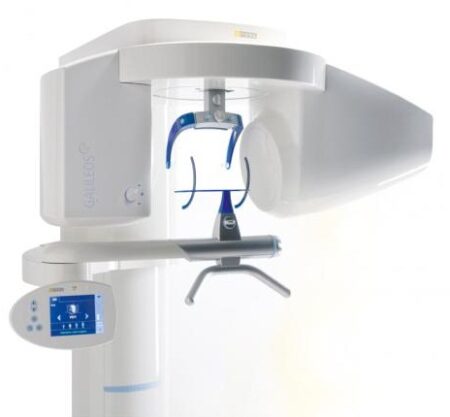
Add cephalometric imaging abilities to your 9000 3D System for even more abilities. Through our revolutionary one-shot technology, cephalometric image acquisition takes less than a second—thereby reducing exposure time and the risk of retakes due to patient movement. With the 9000C 3D System, you can perform a wider range of diagnoses and treatments right in your office, rather than referring patients to imaging centers. Reduce multiple visits, prevent rescheduling issues, and enhance patient care with three versatile imaging abilities right at your fingertips: panoramic, cephalometric, and 3D imaging.Superb image quality, all three modalitiesVariable focal trough adapts to patient’s jaw morphology – generating high-quality, true panoramic images Best-in-class cephalometric image quality with reduced risk of motion blur Ultra-high resolution, low-dose, focused-field CBCT images provide superior visualization of fine bony anatomy, such as radicular structures
Optimized for radiology workflowsDR technology reduces exam time and eliminates steps, such as film processing and cassette handling One-shot cephalometric technology captures images in less than a second, reducing exam time and risk of retakes Dedicated sensors for each modality save technologist time, reduce risk of breakage DICOM conformance ensures easy integration with PACS, RIS, and printing systems
Simple by design Kodak 9000 3D Extraoral Imaging SystemStreamlined user interface, preset programs and face-to-face positioning makes exams quick and simple Accommodates patients of all shapes and sizes and wheelchair accessible Intuitive, easy-to-use imaging software with improved features for reviewing, reporting and sharing images Includes a comprehensive implant planning model to help referring dentists identify exact measurements and create simulations of proposed treatments
A trusted company, proven productsProven product reliability – even in demanding, high-volume environments Broad range of Carestream equipment and professional services such equipment service agreements, site planning and DICOM integration services. - One-stop shopping for digital imaging and IT solutions from a company with over a century of medical and dental imaging expertise
| Panoramic Modality | Cephalometric Modality | Cone Beam CT Modality | Technology CCD – Optical | CCD – Optical Fiber Sensor | One-Shot with CCD Sensor with Optical Fiber | Cone Beam with CMOS Sensor with Optical Fiber | Gray Scale | 14 bit | 14 bit | 15 bit | 3D Field-of-View | n/a | n/a | • Single Volume: 5 cm x 3.75 cm • Stitched Volumes (up to 3): 9 cm x 7 cm x 3.75 cm | Exam Options | • Panoramic • Child Mode • Segmented Panoramic • Maxillary Sinus • LA TMJ x 2 • LA TMJ x 4 | • Cephalometric • Lateral • Oblique • Frontal (AP/PA) • Submento-Vertex • Carpus • Multiple Cephalometric Field Choices: 30 x 30, 24 x 30, 24 x 24, 18 x 24, 18 x 18 | • CBCT • Resolution Options: 76, 100, or 200 micrometers • Location of Exam: User-defined (mandible, maxilla, TMJ) | Input Voltage | 230/240 V – 60 Hz | 230/240 V – 60 Hz | 230/240 V – 60 Hz | Tube Voltage | 60-90 kV (max) | 60-90 kV (max) | 60-90 kV (max) | Tube Current | 2-15 mA (max | 2-15 mA (max | 2-15 mA (max | Frequency | 140 kHZ (max) | 140 kHZ (max) | 140 kHZ (max) | Total Filtration | > 3.5 mm eq. AL | > 3.5 mm eq. AL | > 3.5 mm eq. AL | Weight | 353 lb (160 kg) | 438 lb (199 kg) | 353 lb (160 kg) |
| Schick CDR PanX Laser
FeaturesCDRPanX features proven Schick CDR digital imaging technology and renders crisp, clinically accurate images
Only slightly more expensive than most film-based systems, CDRPanX eliminates costly panoramic film and processing – increasing your economic efficiency
Eight different imaging modes and three laser alignment beams for optimal exposures
Superior vertical travel allows it to be used with patients of any height, including those in wheelchairs
Patient Positioning: 3 laser alignment beams (mid-sagittal, Frankfort, focal trough) bite guide, chin rest, optional temple support
SpecificationsGenerator: Multipulse DC
X-Ray Tube: OCX105
Focal spot size: 0.5 IEC 336
Minimum total filtration: 2.5 mm Al
Anode Voltage: 60-86kV (2kV increments)
Anode Current: 4, 5, 6.3, 8, and 10mA
Exposure time: 19s – adult panoramic
Power Supply: 115V, 50/60Hz
Fusing: T8A
Weight: 474lbs
Electrical Safety Classification: Class 1, type B
Imaging modes (Exposure times): adult (19s), child (15s), left half (10s), right half (10s), anterior (8s), TMJ opened and closed (4x4s), frontal sinuses (8s)
Patient Positioning: 3 laser alignment beams (mid-sagittal, Frankfort, focal trough) bite guide, chin rest, optional temple supportFeatures- CDRPanX features proven Schick CDR digital imaging technology and renders crisp, clinically accurate images
- Only slightly more expensive than most film-based systems, CDRPanX eliminates costly panoramic film and processing – increasing your economic efficiency
- Eight different imaging modes and three laser alignment beams for optimal exposures
- Superior vertical travel allows it to be used with patients of any height, including those in wheelchairs
- Patient Positioning: 3 laser alignment beams (mid-sagittal, Frankfort, focal trough) bite guide, chin rest, optional temple support
Specifications Schick CDR PanX Laser| Generator: | Multipulse DC | | X-Ray Tube: | OCX105 | | Focal spot size: | 0.5 IEC 336 | | Minimum total filtration: | 2.5 mm Al | | Anode Voltage: | 60-86kV (2kV increments) | | Anode Current: | 4, 5, 6.3, 8, and 10mA | | Exposure time: | 19s – adult panoramic | | Power Supply: | 115V, 50/60Hz | | Fusing: | T8A | | Weight: | 474lbs | | Electrical Safety Classification: | Class 1, type B | | Imaging modes (Exposure times): | adult (19s), child (15s), left half (10s), right half (10s), anterior (8s), TMJ opened and closed (4x4s), frontal sinuses (8s) | | Patient Positioning: | 3 laser alignment beams (mid-sagittal, Frankfort, focal trough) bite guide, chin rest, optional temple support |
| 3shape D710 Dental Scanner3Shape D710 Scanner series can scan gypsum and impression versions. The series scanners can integrate into the clinic with minimum staff training. The scanners provide greater practice productivity, a comprehensive list of signals, and the most extensive 3D scans.Where flexibility and productivity are the key standards 3Shape chain scanners are for medium to large labs.The 3Shape D710 is optimized for scanning both the impression and gypsum models and enables high throughput for both advanced and standard indications. These innovative scanners are so easy to use that staff training is barely required. Item fixation is fast, and the scan starts with a click on the program.3SHAPE D710 For Sale3Shape D700 Scanner is both a scanner and impression scanner in 1 unit. It utilizes three measurement movement and two cameras a laser and creates another pass to especially hone. Just one device can be scanned by the apparatus in under 30 seconds and a whole model in about a minute with a degree of accuracy of 20 microns.Features:
-3-axis motion system
-Red-laser Tech
-Gypsum Version scanning
-Impression scanning Alternative
-Multi-die scanningBenefits:
-Easy operation, extreme Precision, and Rapid Scan
-Enables impressions, deep inlays, and Complete undercuts to scan Efficiently
-Supply labs Using ultra-efficient batches and multi-case processingHigh-speed scanning
The D710 is optimized for scanning gypsum and impression versions and enables high throughput for both advanced and standard indications3Shape – leading impression scanning technologies
3Shape’s patented adaptive scanning technology, combined with the scanner’s 3-axis motion and 2 cameras with reduced angle, facilitate the unique impression scanning capability.Adaptive scanning intelligently detects incomplete areas and automatically builds
adaptive scan sequences for capturing full object geometry. | SuniRay 2 Digital Intraoral Sensor Size 1 and Size 2
X-Ray Imaging Properties- Resolution: The pixel resolution of the high resolution (HiRes) sensors exceeds 15 lp/mm. The standard resolution of an X-Ray image, as measured by the modulation transfer function (MTF) exceeds 12 lp/mm. This is measured by using a standard 60 kV intraoral X-ray source.
- Does Efficiency: The sensor will produce a high quality image with an X-Ray dose that is only a fraction of the standard dose required by Dental X-Ray film.
- Diagnostic Efficacy: The system will produce a superior image quality of images obtained when using standard dental X-Ray film. This allows the dentist to diagnose standard intraoral pathologies encountered during screening procedures.
- Wide dynamic range: The sensor pixel well capacity is very high, allowing a higher accommodation of greyscale (bone density) dynamic range.
Sensor Material Biocompatibility SpecificationsThe sensor’s body material and cable material are biocompatible. The sensors and all component materials have been tested to comply with ISO standards, and specifically, the sensor complies with EN IS0-10993 (Biological Evaluation of Medical Devices). | Technical data | Size 1 | Size 2 |
|---|
| Sensor Dimensions (mm) | 39.5 x 26 | 43.5 x 31.5 |
|---|
| Active Area (mm) | 31.1 x 20.2 | 35.2 x 26.2 |
|---|
| Sensor Technology | CMOS APS Fiber Plate | CMOS APS Fiber Plate |
|---|
| Maximum Gray Levels | 4096 | 4096 |
|---|
| Sensor Cable | 3 feet (1m) | 3 feet (1m) |
|---|
| Cable Attachment | Reinforced Strain Relief | Reinforced Strain Relief |
|---|
| USB Module | Integrated USB 2.0 Module | Integrated USB 2.0 Module |
|---|
| Software | Prof. Suni | Prof. Suni |
|---|
X-Ray Imaging Properties SuniRay 2 Digital Intraoral Sensor Size 1 and Size 2- Resolution: The pixel resolution of the high resolution (HiRes) sensors exceeds 15 lp/mm. The standard resolution of an X-Ray image, as measured by the modulation transfer function (MTF) exceeds 12 lp/mm. This is measured by using a standard 60 kV intraoral X-ray source.
- Does Efficiency: The sensor will produce a high quality image with an X-Ray dose that is only a fraction of the standard dose required by Dental X-Ray film.
- Diagnostic Efficacy: The system will produce a superior image quality of images obtained when using standard dental X-Ray film. This allows the dentist to diagnose standard intraoral pathologies encountered during screening procedures.
- Wide dynamic range: The sensor pixel well capacity is very high, allowing a higher accommodation of greyscale (bone density) dynamic range.
|




Reviews
There are no reviews yet.