Due to technological advances with the optical plate and scintillator, all size sensors have increased “sensitivity,” which allows practitioners to decrease the radiation dose €” up to 10% decrease for sizes 1 & 2 and 40% decrease for size 0 (depending on type and age of x-ray generator). In addition, the sensor’s rounded corners and ergonomic design helps make positioning easier than ever.
Other people want this. 15 people have this in their carts right now.
Carestream Kodak Rvg 6100 Digital Xray Sensor Size 1 And Size 2
The RVG 6100 System features a new size 0, 1, 2 sensor for pediatric specialties. The sensor is designed with rounded corners and special bite blocks for patient comfort. All three sensor sizes have new casings with the cable attached to the back of the sensor. New sensor casings for the sensors — size 1 has rounded edges and size 2 has clipped corners help provide a more comfortable experience for the patient, remove the “dead zone” factor, and allow for vertical placement.
RVG 6100 System’s Sensor Features
True Image Resolution
Size 0 : 14 lp/mm
Size 1 : Greater than 20 lp/mm
Size 2 : Greater than 20 lp/mm
Outside Dimensions
Size 0 : 22.2 x 30.8 mm
Size 1 : 27.5 x 37.7 mm
Size 2 : 32.2 x 44.1 mm
Dimensions of Active Area
Size 0 : 17 x 22 mm
Size 1 : 22 x 30 mm
Size 2 : 27 x 36 mm
Matrix Dimensions
Size 0 : 800 x 1200 pixels
Size 1 : 1200 x 1600 pixels
Size 2 : 1440 x 1920 pixels
Highest Quality. Highest Resolution.
The Kodak Carestream RVG 6100 Digital Radiography System has the highest resolution digital imaging sensor on the market today. We understand how image resolution is related to confident diagnoses – and ultimately to the quality of patient care. The RVG 6100 System provides the high resolution you need for complex examinations and to make diagnoses quickly and confidently.
Comfortable and Durable.Carestream Kodak Rvg 6100 Digital Xray
Due to technological advances with the optical plate and scintillator, all size sensors have increased “sensitivity,” which allows practitioners to decrease the radiation dose €” up to 10% decrease for sizes 1 & 2 and 40% decrease for size 0 (depending on type and age of x-ray generator). In addition, the sensor’s rounded corners and ergonomic design helps make positioning easier than ever.
Reviews (0)
Reviews
There are no reviews yet.
Be the first to review “Carestream Kodak Rvg 6100 Digital Xray” Cancel reply
Comfortable and Durable.Due to technological advances with the optical plate and scintillator, all size sensors have increased "sensitivity," which allows practitioners to decrease the radiation dose €” up to 10% decrease for sizes 1 & 2 and 40% decrease for size 0 (depending on type and age of x-ray generator). In addition, the sensor's rounded corners and ergonomic design helps make positioning easier than ever.
Milling Unit:• Choose from CEREC MC, CEREC MC X or CEREC MC XL
• Get access to the entire range of chairside treatment with up to 40 mm block size, including bridges and abutments
• Create fully anatomic, precise treatments quickly
• CEREC Guide 2 surgical guide
• Dry mill ready
• Includes CEREC software
iTero HD2.9 simply allows to digitally scan a prepared tooth and wirelessly send this information directly to the dental laboratory for fabrication of a CAD/CAM crown without the gooey mess.
Schick Technologies has brought digital panoramic imaging to a whole new level of affordability and simplicity.
3 Shape D2000 Dental lab Scanner Optimized processing speed: creates faster performance and better scanning workflows for several indications Shortens waiting time between creating orders and starting the scan, and enables technicians to begin next scan without having to wait for post processing.
The DEXIS Digital X-ray System includes a highend CCD sensor, portable PC card and DEXusb connection options, DEXIS imaging software, a complete set of sensor holders and accessories, on-site training, a printed instruction manual, training tutorials on CD, a quick reference guide for the operatory, and 1 year of DEXcare support.
Content
Carestream Kodak Rvg 6100 Digital Xray Sensor Size 1 And Size 2The RVG 6100 System features a new size 0, 1, 2 sensor for pediatric specialties. The sensor is designed with rounded corners and special bite blocks for patient comfort. All three sensor sizes have new casings with the cable attached to the back of the sensor. New sensor casings for the sensors — size 1 has rounded edges and size 2 has clipped corners help provide a more comfortable experience for the patient, remove the “dead zone” factor, and allow for vertical placement.RVG 6100 System’s Sensor Features
True Image Resolution
Size 0 : 14 lp/mm
Size 1 : Greater than 20 lp/mm
Size 2 : Greater than 20 lp/mmConnection : USB 2 – High SpeedTechnology : KODAK SensorPurpose
Size 0 : Pediatric Exams
Size 1 : General Purpose Sensor
Size 2 : Bitewing RadiographsOutside Dimensions
Size 0 : 22.2 x 30.8 mm
Size 1 : 27.5 x 37.7 mm
Size 2 : 32.2 x 44.1 mmDimensions of Active Area
Size 0 : 17 x 22 mm
Size 1 : 22 x 30 mm
Size 2 : 27 x 36 mmMatrix Dimensions
Size 0 : 800 x 1200 pixels
Size 1 : 1200 x 1600 pixels
Size 2 : 1440 x 1920 pixelsHighest Quality. Highest Resolution.The Kodak Carestream RVG 6100 Digital Radiography System has the highest resolution digital imaging sensor on the market today. We understand how image resolution is related to confident diagnoses - and ultimately to the quality of patient care. The RVG 6100 System provides the high resolution you need for complex examinations and to make diagnoses quickly and confidently.Comfortable and Durable.Carestream Kodak Rvg 6100 Digital XrayDue to technological advances with the optical plate and scintillator, all size sensors have increased "sensitivity," which allows practitioners to decrease the radiation dose €” up to 10% decrease for sizes 1 & 2 and 40% decrease for size 0 (depending on type and age of x-ray generator). In addition, the sensor's rounded corners and ergonomic design helps make positioning easier than ever.
CEREC Primescan AC Intraoral scanner
Cerec Primescan from Dentsply Sirona is the most advanced digital scanner on the market. It’s providing a level of accuracy never before seen in dentistry. At the peak of innovation, it is widening the gap between those with and without digital dentistry. The Prime Scan is the fastest most accurate intraoral scanner to date. With new scanning technology, it is the ultimate in scanning. Prime Scan and CEREC 5.0 combinations are greatly reduced because more data allows many applications to become automated.For more than 30 years, CEREC® has provided technological precision, superior design and excellent performance to thousands of practices. Now, experience a brand-new fully integrated solution with CEREC Primescan AC. Acquire precise digital impressions with Primescan, design your proposal on the Primescan AC using the intelligent software, then produce precise, smooth restorations with your choice of milling unit. Find the next level of productivity and patient care – it’s all here.How Does CEREC Primescan Work?
CEREC Primescan is an alternative to traditional intraoral dental impressions, which use a putty-like substance to take physical impressions of your teeth. This method does not allow for digital assessment of your mouth, and some patients find it the process of physical impressions to be uncomfortable.CEREC Primescan uses a wand-like device, loaded with powerful imaging sensors which can capture more than 50,000 images of your mouth per second. These images are quickly stitched and compiled into a 3D image of your teeth, jaw, gums, and mouth, and displayed on a high-definition screen for immediate assessment by our doctors.Using this sensor, our doctors can image your entire mouth up to 20mm in depth, including gums, blood, and bone structures, quickly and accurately. The Primescan software (as compared to the Omnicam) creates high-resolution 3D models to assess your oral health and create the very best crowns, implants, removable prosthesis, restorations, and even 3D prints. With just a few quick sweeps of the CEREC Primescan device, we’ll be able to generate an in-depth, modifiable 3D image of your mouth in minutes.The Benefits of CEREC Primescan:
An excellent choice for outstanding results: Primescan is your perfect starting point in digital dentistry. No matter how you would like to design your workflows, Primescan is the enabler for efficient digital workflows – both chairside in your practice and with your preferred partners.Well designed and perfectly compatible: Along with the high-performance intraoral scanner comes the new Acquisition Center. Together, they form a highly effective and most comfortable system – meeting your needs with two individual software configurations:So, why is CEREC Primescan such a big deal? Here are just a few improvements to this sophisticated system:Shorter wait times – We can quickly image your mouth without waiting for dental impressions to set, allowing us to treat each patient more quickly and effectively. A full jaw impression only takes 2 minutes. This adds up to less time spent in the dental chair.Real-time image processing – Our dentists can quickly adjust and modify scanned images using filters and other tools, leading to more accurate diagnostics and better overall patient outcomes.Extremely versatile – CEREC Primescan can be used for almost any dental procedure. From implant dentistry to the creation of CEREC same-day crowns, the straightening of teeth, whitening, development of dentures and partial dentures.CEREC Primescan AC with CEREC Software Sirona Advantages:
Enables all kinds of treatment, from a single tooth to full arch
With Primescan intraoral scanning is now more accurate, faster and easier
Allows an efficient workflow, both chairside and with preferred partners
Along with the high-performance intraoral scanner comes the new Acquisition Center, together forming an effective and most comfortable system, meeting your needs with two individual software configurations: Primescan AC with Connect Software (supports the connection to established partners workflows) and CEREC Primescan AC with CEREC Software (ideal for cabinets)
Smart Pixel Sensor technology – allows superior precision and excellent detail reproduction, processes more than 1 million 3D points per second, producing photorealistic and highly accurate data
Due to its ability to scan at an incredible data density, it delivers complete 3D structures of everything in its field of view
Its dynamic depth scan technology enables perfect sharpness and outstanding precision, even at a measuring depth up to 20 mm
Easy to use
Easy scanning of all dental materials and hard to access areas
Allows you to start scanning right away
Self-heating for fog-free scanning
The increased field of view visualizes larger areas with fewer sweeps and immediate precision
Faster scanning with a smooth scan flow
Consolidating more than 50.000 images per second and fast processing of exactly the data the software needs Easy capturing and quicker processing of more data in higher resolution
Complete 3D-scan models are displayed immediately, no matter how fast you scan
Accelerate your workflow: Due to seamless, validated and open data transfer options, labs and other third parties are supplied with high-resolution models at an instant
3 different sleeves (disposable and stainless steel, with sapphire glass or autoclavable)
Acquisition Center – Smart features and greater comfort: intuitive use via movable 16:9 touchscreen and touchpad for perfect ergonomics
Kinematics for perfect ergonomic positioning
Smart Hygiene concept for fast and easy disinfection
Full mobility with more than 60-min. battery buffer
CEREC Primescan AC with CEREC Software – complete chairside system, which offers flexible data export options, with automatized workflow thanks to Artificial Intelligence and touch-enabled and intuitive user interface
Milling Unit:• Choose from CEREC MC, CEREC MC X or CEREC MC XL
• Get access to the entire range of chairside treatment with up to 40 mm block size, including bridges and abutments
• Create fully anatomic, precise treatments quickly
• CEREC Guide 2 surgical guide
• Dry mill ready
• Includes CEREC softwareStriking in every detail: The new Acquisition CenterThe new Acquisition Center is a work station designed for modern dentistry and with the dentist in mind. It comes with a touchpad and a 16:9 wide-format movable touchscreen, offering you a highly intuitive and ergonomic work platform.Your benefits at a glance:
Touchscreen & Touchpad for most comfortable, intuitive use
Kinematics for perfect ergonomic positioning
Smart Hygiene concept for fast and easy disinfection
Mobility concept for full mobility with more than 60-min. battery buffer
The best choice for your practice: Take digital impressions and continue working with your trusted partners or enjoy the benefits of complete chairside workflows.Primescan meets your needs with two individual software configurations:Primescan AC with Connect Software:
Supports data transfer options to your preferred partners
Secure and encrypted data transfer through Connect Case Center Inbox
Easy upgradability to full chairside workflow
Touch-enabled and intuitive user interface
CEREC Primescan AC with CEREC Software:
Supports full chairside workflows for single-visit dentistry
Flexible data export options
Automatized workflow thanks to Artifical Intelligence
Touch-enabled and intuitive user interface
Technical Data:CEREC Primescan AC Intraoral scanner
CAD system for high-precision intraoral optical impressions
High-resolution, heated intraoral 3D scanner
Integrated image processing
High processing power due to state-of-the-art processor
Trackball or touchpad
Hand and foot controlled enter keys
Ethernet port and WLAN
USB interfaces
The high-resolution 3D intraoral scanner allows image acquisition with the image control inside the scanner, the image data transfer is made through USB 2.0 interface
21.5″ inch TFT LED flat-screen display, HD resolution: 1920 x 1080 pixels
Mobile housing, with no water or air connection required
Voltage: 100 – 240 VAC/50-60Hz
Nominal current: 5.0 – 2.1 A
Type of protection against electric shock: Class I device
iTero HD2.9 simply allows to digitally scan a prepared tooth and wirelessly send this information directly to the dental laboratory for fabrication of a CAD/CAM crown – without the gooey mess. The wireless transmission saves shipping time and eliminates distortion that can often be found with traditional stone modelsThe digital scan is, quite literally, digitally perfect. We then deliver the digital scan to our lab where a precision fitting restoration is created.iTero HD2.9 system eliminated the uncomfortable patient experience of traditional impressions, utilizes advanced state of the art digital technology, and provides with superior fitting restorations with little or no adjustmentsiTero Intra-Oral Scanner
Model: HD2.9
2014 Date of Mfg.
Brochure/Video: Click Here
Limited Use
Includes:
iTero Software (pre-loaded)
90 Day Parts Warranty
User Manual(s)
Free Standard Shipping (Commercial USA Addresses)
Its ergonomic design comes with safe docking station. It has thin fiber optic cable which enables quick data transfer from the wand to the cart. This optimized cable length assists various cart positions in the operatory.Orthodontic treatment is more than just straightening teeth; it’s about ensuring your teeth and jaws are in alignment. To achieve this, we need to get an accurate look at where your teeth are positioned. To do this, we use the iTero Intraoral digital scanner.A good impressionIn the orthodontic and dental industries, ‘impressions’ is the term we use for getting a good picture of where your teeth currently sit. These impressions were previously taken using a type of dental cement. Pasted on your teeth, patients were required to lie for a few minutes while the cement-like material set. The dental professional could then remove the set material which had formed an ‘impression’ of your teeth.This process was messy, rather unpleasant and not very comfortable. Fortunately, that method is a thing of the past. These days, we use scanning technology and at L&L Orthodontics, our technology of choice is the iTero Intraoral digital scanner.
What is the iTero scanner?
The iTero scanner is new 3D orthodontic scanning technology. Using the latest developments in intraoral scanning, the iTero scanner accurately scans your teeth as part of your orthodontic assessment. The iTero scanner is made up of a small, wand-like device which is used to scan your teeth and jaws. The data it is collecting then appears on the computer screen, allowing you to see the ‘impressions’ the scanner is taking.The iTero scanner works in with Invisalign®* treatment and lets you see what your teeth will look like post-treatment, even before you’ve started. Before we recommend a course of treatment for you, we’ll take impressions of your teeth using the iTero scanner to ensure we are making our recommendation on accurate information.To find out more about the iTero scanner and other orthodontic technology we use here at L&L Orthodontics, book your specialist consultation today.
Schick CDR PanX Laser
FeaturesCDRPanX features proven Schick CDR digital imaging technology and renders crisp, clinically accurate images
Only slightly more expensive than most film-based systems, CDRPanX eliminates costly panoramic film and processing – increasing your economic efficiency
Eight different imaging modes and three laser alignment beams for optimal exposures
Superior vertical travel allows it to be used with patients of any height, including those in wheelchairs
Patient Positioning: 3 laser alignment beams (mid-sagittal, Frankfort, focal trough) bite guide, chin rest, optional temple support
SpecificationsGenerator: Multipulse DC
X-Ray Tube: OCX105
Focal spot size: 0.5 IEC 336
Minimum total filtration: 2.5 mm Al
Anode Voltage: 60-86kV (2kV increments)
Anode Current: 4, 5, 6.3, 8, and 10mA
Exposure time: 19s – adult panoramic
Power Supply: 115V, 50/60Hz
Fusing: T8A
Weight: 474lbs
Electrical Safety Classification: Class 1, type B
Imaging modes (Exposure times): adult (19s), child (15s), left half (10s), right half (10s), anterior (8s), TMJ opened and closed (4x4s), frontal sinuses (8s)
Patient Positioning: 3 laser alignment beams (mid-sagittal, Frankfort, focal trough) bite guide, chin rest, optional temple supportFeatures
CDRPanX features proven Schick CDR digital imaging technology and renders crisp, clinically accurate images
Only slightly more expensive than most film-based systems, CDRPanX eliminates costly panoramic film and processing – increasing your economic efficiency
Eight different imaging modes and three laser alignment beams for optimal exposures
Superior vertical travel allows it to be used with patients of any height, including those in wheelchairs
– Impression scanning:
Multi line scanning in the D2000 and D1000 improves coverage and performance for accurate scanning of impressions to enable restorative design without the need to pour gypsum models.
All in One scanning of triple tray impressions Special mount enables triple-tray scanning capture of upper, lower and bite in one scan.– Improved workflows for single tray impressions:
Provides support for single tray impression scanning including bite alignment.The software upgrades are available for all relevant 3Shape lab scanner models. Current lab scanner owners will receive the upgrades as a LABcare software update, where applicable.Impression scanning the future is now:
The D2000 and the smaller D1000 lab scanner feature multi-line technology. Multi-line technology utilizes multiple scan lines in a single shot to significantly improve cavity and impression capture.This enables the D2000 to accurately scan analog impressions sent to the lab by a dentist. This in turn, eliminates the need for labs having to pour or create a gypsum model from the impression. This saves several steps in the workflow and allows labs to print instead, cost effective 3D models derived from the analog impression if they need a physical working model.While all 3shape scanners support impression scanning only the D2000 and D1000 feature multi line technology.“Impression scanning is today’s reality and the future. No waiting for models to set, no plaster inaccuracies and we reduce costs. Every lab should invest in this technology,” Carlo Backes, Excent Tandtechniek BV, Holland.About the D2000 dental lab scanner:
Introduced at the 2015 Midwinter show, the D2000 was named a top 10 coolest product at the event. The D2000 captivated the industry by having room for two models in its interior, enabling technicians to scan two models at once and perform a complete case like a three-unit bridge in just one scan.D2000 All in-one scanning for max volume throughputs
– 4 x 5 MP cameras, Blue LED Multi-line
– Accuracy: 5 µm (ISO 12836) / 8 µm (Implant bar)
– Die scan time: 15 sec
– Full arch scan time: 20 sec
– Color texture scanning
– No need for separate die scan
– All in One scanning: room for 2 models
– Impression scanningIncludes:3SHAPE D2000 DENTAL LAB 3D SCANNER
– Remainder of a 5 year factory warranty
– 3 Shape Dental Designer w/Additional Seat
– 3 Shape Model Builder
– 3 Shape Occlusion Set Up Tool
– ScanIt Impression Module
– Impression Fixture
– Anatomical CAD Library
– Calibration Kit
– HP Envy Phoenix Desktop PC
Dexis Intraoral Digital Dental X-Ray Sensor
Software Ease of Use
According to the manufacturer, the DEXIS x-ray software was designed to be highly effective throughout diagnosis, treatment planning, and treatment. It is intended to serve as an imaging hub, managing all digital images, including intra- and extraoral radiographs and photographs. The manufacturer states that the images can be quickly organized, stored, retrieved, printed, and shared with patients and colleagues. The 9 dentists who participated in this product evaluation were asked to rate the DEXIS Digital Imaging software on its ease of use with regard to taking and accessing radiographs and for diagnosis, patient education, and communication with colleagues and insurance companies. Six of the evaluators rated the system as excellent and 3 rated it as very good. One evaluator commented that “when communicating with patients, we use DEXIS images displayed on a monitor in front of the patient every day on virtually every patient.”Software Integration
The manufacturer claims that the DEXIS imaging software can be easily integrated with all major practice management systems. According to DEXIS, the DEXIS Integrator automatically synchronizes with the patient record.
When the evaluators were asked to rate the software integration with practice management software and interface with hardware (eg, cameras, panoramics, and scanners), 78% rated it as excellent to very good, while 22% rated it as good. One of the evaluators said, “The bridge to our practice management systems is seamless. We use [the] DEXIS digital pan[oramic] so that we can store panoramic x-rays easily in the same location as full series. We use an intraoral camera to store preoperative and interoperative photographs for excellent documentation.”Sensor Comfort and Ease of Use
According to DEXIS, the heart of their digital system is the PerfectSize sensor. The manufacturer states that their single-size sensor has 3 advantages:
1) because the electronics are on the back of the sensor, the active imaging area is most of the total sensor size;
2) the beveled corners, slim profile, and rounded casing allow for accurate placement and patient comfort;
3) patented cord placement allows the practitioner to take vertical and horizontal bitewings and all periapicals with only 1 universally sized sensor. The evaluators were asked to rate both the ease of use of the sensor and patient comfort. Five evaluators rated ease of use excellent and 4 rated it very good. With regard to patient comfort, 3 evaluators rated the sensor excellent and 6 rated it very good. One evaluator noted that he experienced a few issues when placing the sensor in some children and patients with very small mouths. Another evaluator, however, is very happy with the sensor, saying, “I chose DEXIS to take a full radiographic series on every patient. It was the only direct image system we tested that met that objective.”Image Quality Dexis Intraoral Digital Dental X-Ray Sensor
According to DEXIS, their digital xray system provides detailed image quality and includes image enhancement tools that increase diagnostic capabilities. When the 9 evaluators were asked to rate the system’s image quality and enhancement aids, 89% rated them as excellent to very good. One of the evaluators commented that the system’s “ClearVu and image magnification allow us to diagnose very effectively.”Overall Satisfaction
When asked to rate their overall satisfaction with the DEXIS Digital X-ray System, 89% of the evaluators rated it as excellent to very good. Commented one evaluator, “DEXIS has been very responsive to our needs.”


 An excellent choice for outstanding results: Primescan is your perfect starting point in digital dentistry. No matter how you would like to design your workflows, Primescan is the enabler for efficient digital workflows – both chairside in your practice and with your preferred partners.Well designed and perfectly compatible: Along with the high-performance intraoral scanner comes the new Acquisition Center. Together, they form a highly effective and most comfortable system – meeting your needs with two individual software configurations:So, why is CEREC Primescan such a big deal? Here are just a few improvements to this sophisticated system:Shorter wait times – We can quickly image your mouth without waiting for dental impressions to set, allowing us to treat each patient more quickly and effectively. A full jaw impression only takes 2 minutes. This adds up to less time spent in the dental chair.Real-time image processing – Our dentists can quickly adjust and modify scanned images using filters and other tools, leading to more accurate diagnostics and better overall patient outcomes.Extremely versatile – CEREC Primescan can be used for almost any dental procedure. From implant dentistry to the creation of CEREC same-day crowns, the straightening of teeth, whitening, development of dentures and partial dentures.CEREC Primescan AC with CEREC Software Sirona Advantages:
 The new Acquisition Center is a work station designed for modern dentistry and with the dentist in mind. It comes with a touchpad and a 16:9 wide-format movable touchscreen, offering you a highly intuitive and ergonomic work platform.Your benefits at a glance:
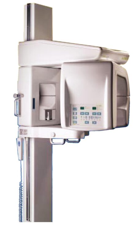


Reviews
There are no reviews yet.